Skip to Content
MilliporeSigma
All Photos(1)

Documents

927651

Sigma-Aldrich

TissueFab® bioink kit

(Gel)ma Fibrin (UV/365), low endotoxin

Synonym(s):

Fibrin, Fibrinogen, GelMA, Gelatin methacrylamide, Gelatin methacrylate, Gelatin methacryloyl, Thrombin

Sign Into View Organizational & Contract Pricing


About This Item

UNSPSC Code:
12352201
NACRES:
NA.21

description

HNMR in D2O at 40°C

Quality Level

form

(Solid chunks, fibers or powder)

impurities

<10 CFU/g Bioburden (Fungal)
<10 CFU/g Bioburden (Total Aerobic)
<125 EU/g Endotoxin

color

white

storage temp.

2-8°C

Looking for similar products? Visit Product Comparison Guide

General description

Gelatin methacryloyl (GelMA) is a polymerizable hydrogel material derived from natural extracellular matrix (ECM) components. Due to its low cost, abundance, and retention of natural cell binding motifs, gelatin has become a highly sought material for tissue engineering applications. Fibrinogen is a soluble glycoprotein produced by liver. It can be enzymatically polymerized to fibrin monomers by thrombin and then to fibrin based blood clot during blood coagulation. With multi cell binding sites, such as RGD (Arg-Gly-Asp), fibrin plays an important role in cellular-matrix interaction, can promote cell adhesion, proliferation and migration.

Application

The addition of photocrosslinkable methacrylamide functional groups in GelMA allows the synthesis of biocompatible, biodegradable, and non-immunogenic hydrogels that are stable in biologically relevant conditions and promote cell adhesion, spreading, and proliferation. Gelatin methacrylate based bioinks have been used to bioprint osteogenic , chondrogenic , hepatic , adipogenic , vasculogenic , epithelial , endothelial , cardiac valve , skin , tumor and other tissues and constructs. Fibrinogen and fibrin has been widely utilized in tissue engineering for wound healing, neural regeneration, bone generation and vascularization, etc.
TissueFab® bioink kit- (Gel)ma Fibrin (UV/365), low endotoxin is a ready-to-use bioink which is formulated for high cell viability, osteoinduction and printing fidelity and is designed for extrusion-based 3D bioprinting and subsequent crosslinking with exposure to 405nm visible light. TissueFab® bioink kit- (Gel)ma Fibrin (UV/365), low endotoxin can be used with most extrusion-based bioprinters, are biodegradable, and are compatible with human dermal fibroblasts (HDFs). TissueFab® bioink kit- (Gel)ma Fibrin (UV/365), low endotoxin enables the precise fabrication of osteogenic 3D cell models and tissue constructs for research in 3D cell biology, tissue engineering, in vitro tissue models, and regenerative medicine.

The protocol can be found under "More Documents" at the bottom of the page.

TissueFab® bioink kit- (Gel)ma Fibrin (UV/365), low endotoxin contains:
2- 500 mg lyophilized ink components
1- lyophilized thrombin powder
1- 10 ml HEPES buffer.

Features and Benefits

In addition to fast gelation, the methacrylamide functional group can also be used to control the hydrogel physical parameters such as pore size, degradation rate, and swell ratio. Temporal and spatial control of the crosslinking reaction can be obtained by adjusting the degree of functionalization and polymerization conditions, allowing for the fabrication of hydrogels with unique patterns, 3D structures, and morphologies.

Low Endotoxin, low bioburden: Endotoxins have been demonstrated negatively impact cellular growth, morphology, differentiation, inflammation and protein expression. Bioburden is defined as the number of contaminated organisms found in a given amount of material. We test each lot for endotoxins as well as total bioburden (aerobic and fungal) to minimize unwanted interactions. For more information: https://www.sigmaaldrich.com/US/en/technical-documents/technical-article/microbiological-testing/pyrogen-testing/what-is-endotoxin

Legal Information

TISSUEFAB is a registered trademark of Merck KGaA, Darmstadt, Germany

related product

Product No.
Description
Pricing

pictograms

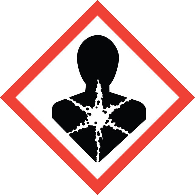
Health hazardExclamation mark

signalword

Danger

Hazard Classifications

Eye Irrit. 2 - Resp. Sens. 1 - Skin Irrit. 2 - STOT SE 3

target_organs

Respiratory system

wgk_germany

WGK 3

flash_point_f

Not applicable

flash_point_c

Not applicable


Certificates of Analysis (COA)

Search for Certificates of Analysis (COA) by entering the products Lot/Batch Number. Lot and Batch Numbers can be found on a product’s label following the words ‘Lot’ or ‘Batch’.

Already Own This Product?

Find documentation for the products that you have recently purchased in the Document Library.

Visit the Document Library

Emily Abelseth et al.
ACS biomaterials science & engineering, 5(1), 234-243 (2019-01-14)
3D bioprinting offers the opportunity to automate the process of tissue engineering, which combines biomaterial scaffolds and cells to generate substitutes for diseased or damaged tissues. These bioprinting methods construct tissue replacements by positioning cells encapsulated in bioinks into specific
N Laurens et al.
Journal of thrombosis and haemostasis : JTH, 4(5), 932-939 (2006-05-13)
Fibrinogen and fibrin play an important role in blood clotting, fibrinolysis, cellular and matrix interactions, inflammation, wound healing, angiogenesis, and neoplasia. The contribution of fibrin(ogen) to these processes largely depends not only on the characteristics of the fibrin(ogen) itself, but
Y Shi et al.
Biomedical materials (Bristol, England), 13(3), 035008-035008 (2018-01-09)
Three-dimensional bioprinting is an emerging technology for fabricating living 3D constructs, and it has shown great promise in tissue engineering. Bioinks are scaffold materials mixed with cells used by 3D bioprinting to form a required cell-laden structure. In this paper
Wanjun Liu et al.
Advanced healthcare materials, 6(12) (2017-05-04)
Bioprinting is an emerging technique for the fabrication of 3D cell-laden constructs. However, the progress for generating a 3D complex physiological microenvironment has been hampered by a lack of advanced cell-responsive bioinks that enable bioprinting with high structural fidelity, particularly
B Duan et al.
Acta biomaterialia, 10(5), 1836-1846 (2013-12-18)
Tissue engineering has great potential to provide a functional de novo living valve replacement, capable of integration with host tissue and growth. Among various valve conduit fabrication techniques, three-dimensional (3-D) bioprinting enables deposition of cells and hydrogels into 3-D constructs

Our team of scientists has experience in all areas of research including Life Science, Material Science, Chemical Synthesis, Chromatography, Analytical and many others.

Contact Technical Service