Skip to Content
MilliporeSigma
All Photos(1)

Documents

04-1488

Sigma-Aldrich

Anti-HIRA Antibody, clone WC119

clone WC119, from mouse

Synonym(s):

DiGeorge critical region gene 1, HIR (histone cell cycle regulation defective) homolog A (S. cerevisiae), HIR histone cell cycle regulation defective homolog A, HIR histone cell cycle regulation defective homolog A (S. cerevisiae), TUP1-like enhancer of

Sign Into View Organizational & Contract Pricing


About This Item

UNSPSC Code:
12352203
eCl@ss:
32160702
NACRES:
NA.41

biological source

mouse

Quality Level

antibody form

purified immunoglobulin

antibody product type

primary antibodies

clone

WC119, monoclonal

species reactivity

mouse

species reactivity (predicted by homology)

human (based on 100% sequence homology)

technique(s)

immunoprecipitation (IP): suitable
western blot: suitable

isotype

IgG1κ

NCBI accession no.

UniProt accession no.

shipped in

wet ice

target post-translational modification

unmodified

Gene Information

human ... HIRA(7290)

General description

HIRA (TUP1 like enhancer of split protein 1) is a protein localized primarily in the nucleus. It functions as a histone chaparone to preferentially place histone H3.3 on nucleosome. It is broadly expressed in a variety of tissues and is expressed during embryogenesis. Data suggests HIRA plays a part in mechanisms of transcriptional regulation similar to that played by yeast HIR1 and HIR2 together. The HIRA protein has been shown to interact with HIRIP3, HIRIP5, histones H2B and H4, and PAX3. HIRA may play a part in the etiology of the DiGeorge syndrome (DGS), a developmental disorder. The clinical features of this disease include absence or hypoplasia of the thymus and parathyroid glands, cardiovascular malformations, facial dysplasia, a cleft palate and mental retardation.

Specificity

This antibody recognizes HIRA.

Immunogen

Epitope: Unknown
GST-tagged recombinant protein corresponding to human HIRA.

Application

Anti-HIRA Antibody, clone WC119 is a Mouse Monoclonal Antibody for detection of HIRA also known as HIR (histone cell cycle regulation defective) homolog & has been validated in WB & IP.
Immunoprecipitation Analysis: A previous lot was used by an independent laboratory in IP. (Hall, C., et al. (2001). Molecular and Cellular Biology. 21(5):1854-1865.)
Research Category
Epigenetics & Nuclear Function
Research Sub Category
Chromatin Biology

Chaperones

Quality

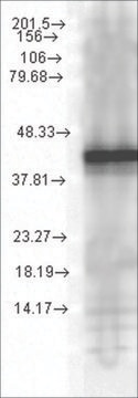
Evaluated by Western Blot in NIH/3T3 cell lysate.

Western Blot Analysis: 0.44 µg/ml of this antibody detected HIRA on 10 µg of NIH/3T3 cell lysate.

Target description

~ 112 kDa

Physical form

Format: Purified
Protein G Purified
Purified mouse monoclonal IgG1κ in buffer containing 0.1 M Tris-Glycine (pH 7.4, 150 mM NaCl) with 0.05% sodium azide.

Storage and Stability

Stable for 1 year at 2-8°C from date of receipt.

Analysis Note

Control
NIH/3T3 cell lysate

Other Notes

Concentration: Please refer to the Certificate of Analysis for the lot-specific concentration.

Disclaimer

Unless otherwise stated in our catalog or other company documentation accompanying the product(s), our products are intended for research use only and are not to be used for any other purpose, which includes but is not limited to, unauthorized commercial uses, in vitro diagnostic uses, ex vivo or in vivo therapeutic uses or any type of consumption or application to humans or animals.

Not finding the right product?  

Try our Product Selector Tool.

wgk_germany

WGK 1

flash_point_f

Not applicable

flash_point_c

Not applicable


Certificates of Analysis (COA)

Search for Certificates of Analysis (COA) by entering the products Lot/Batch Number. Lot and Batch Numbers can be found on a product’s label following the words ‘Lot’ or ‘Batch’.

Already Own This Product?

Find documentation for the products that you have recently purchased in the Document Library.

Visit the Document Library

Tetsuro Komatsu et al.
Journal of virology, 86(12), 6701-6711 (2012-04-13)
In infected cells, the chromatin structure of the adenovirus genome DNA plays critical roles in its genome functions. Previously, we reported that in early phases of infection, incoming viral DNA is associated with both viral core protein VII and cellular
Zhexin Zhu et al.
Cell stem cell, 20(2), 274-289 (2016-12-13)
The chromatin landscape and cellular metabolism both contribute to cell fate determination, but their interplay remains poorly understood. Using genome-wide siRNA screening, we have identified prohibitin (PHB) as an essential factor in self-renewal of human embryonic stem cells (hESCs). Mechanistically, PHB
Raul Bardini Bressan et al.
Cell stem cell, 28(5), 877-893 (2021-02-26)
Point mutations within the histone H3.3 are frequent in aggressive childhood brain tumors known as pediatric high-grade gliomas (pHGGs). Intriguingly, distinct mutations arise in discrete anatomical regions: H3.3-G34R within the forebrain and H3.3-K27M preferentially within the hindbrain. The reasons for
Kristina Ivanauskiene et al.
Genome research, 24(10), 1584-1594 (2014-07-23)
Histone variant H3.3 is deposited in chromatin at active sites, telomeres, and pericentric heterochromatin by distinct chaperones, but the mechanisms of regulation and coordination of chaperone-mediated H3.3 loading remain largely unknown. We show here that the chromatin-associated oncoprotein DEK regulates
Camille Cohen et al.
PLoS pathogens, 14(9), e1007313-e1007313 (2018-09-21)
Herpes simplex virus 1 (HSV-1) latency establishment is tightly controlled by promyelocytic leukemia (PML) nuclear bodies (NBs) (or ND10), although their exact contribution is still elusive. A hallmark of HSV-1 latency is the interaction between latent viral genomes and PML

Our team of scientists has experience in all areas of research including Life Science, Material Science, Chemical Synthesis, Chromatography, Analytical and many others.

Contact Technical Service