Skip to Content
MilliporeSigma
All Photos(2)

Documents

05-1049

Sigma-Aldrich

Anti-Met (extracellular) Antibody, clone 4F8.2

clone 4F8.2, from mouse

Synonym(s):

HGF receptor, HGF/SF receptor, Met proto-oncogene tyrosine kinase, SF receptor, Scatter factor receptor, met proto-oncogene, met proto-oncogene (hepatocyte growth factor receptor), oncogene MET

Sign Into View Organizational & Contract Pricing


About This Item

UNSPSC Code:
12352203
eCl@ss:
32160702
NACRES:
NA.41

biological source

mouse

Quality Level

antibody form

purified antibody

antibody product type

primary antibodies

clone

4F8.2, monoclonal

species reactivity

human

technique(s)

immunohistochemistry: suitable (paraffin)
western blot: suitable

isotype

IgG2bκ

NCBI accession no.

UniProt accession no.

shipped in

wet ice

target post-translational modification

unmodified

Gene Information

human ... MET(4233)

Related Categories

General description

The proto-oncogene Met (c-Met, HGF/SF Receptor, HGFR) is a receptor tyrosine kinase that is activated upon Hepatocyte Growth Factor (HGF) binding. This stimulates the autophosphorylation on Tyr1234/Tyr1235 which leads to pleiotropic downstream signaling events in epithelial cells that stimulate multiple cellular processes including cell proliferation, growth, differentiation, cell migration/ invasion, and tumorigenesis. These effects are mediated by SH2 domain-containing proteins that bind to the C-terminal tail of c-Met. Chronic stimulation of Met on cancer cells is thought to play a role in metastasis and dysregulation of c-Met activity is directly associated with oncogenesis, making it an important cancer target.

Specificity

Detects Met on its extracellular domain.

Immunogen

Epitope: Extracelluar domain (N-term)
GST-fusion protein corresponding to amino acids 80-434 of human Met.

Application

Detect Met (extracellular) using this Anti-Met (extracellular) Antibody, clone 4F8.2 validated for use in IH(P) & WB.
Research Category
Signaling
Research Sub Category
Growth Factors & Receptors
Western Blot Analysis:
This lot detected Met at 1:1,000 dilution in HEK293 cell lysate resolved via SDS-PAGE and transferred to PVDF.

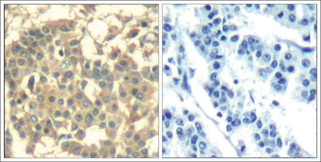
Immunohistochemistry Analysis :

Quality

Routinely evaluated by Western Blot.

Western Blot Analysis:
This lot detected Met at 1:1,000 dilution in HEK293 cell lysate resolved via SDS-PAGE and transferred to PVDF.

Target description

170 kDa

Linkage

Replaces: 05-237

Physical form

Format: Purified
Protein G Purified
Purified mouse monoclonal in 0.1M Tris-Glycine (pH7.4), 150mM NaCl with 0.05% NaN3.

Storage and Stability

Stable for 1 year at 2-8°C from date of receipt.
Handling Recommendations: Upon receipt, and prior to removing the cap, centrifuge the vial and gently mix the solution.

Other Notes

Concentration: Please refer to the Certificate of Analysis for the lot-specific concentration.

Disclaimer

Unless otherwise stated in our catalog or other company documentation accompanying the product(s), our products are intended for research use only and are not to be used for any other purpose, which includes but is not limited to, unauthorized commercial uses, in vitro diagnostic uses, ex vivo or in vivo therapeutic uses or any type of consumption or application to humans or animals.

Not finding the right product?  

Try our Product Selector Tool.

wgk_germany

WGK 1

flash_point_f

Not applicable

flash_point_c

Not applicable


Certificates of Analysis (COA)

Search for Certificates of Analysis (COA) by entering the products Lot/Batch Number. Lot and Batch Numbers can be found on a product’s label following the words ‘Lot’ or ‘Batch’.

Already Own This Product?

Find documentation for the products that you have recently purchased in the Document Library.

Visit the Document Library

Ilia Atanelishvili et al.
PloS one, 11(9), e0162357-e0162357 (2016-09-02)
Pulmonary fibrosis represents the terminal stage of a diverse group of lung diseases including scleroderma associated interstitial lung disease. The molecular mechanisms underlying the pathogenesis of lung fibrosis are not well understood and there is a great need for more
Nolan L Boyd et al.
Tissue engineering. Part A, 19(1-2), 211-223 (2012-09-14)
The microvasculature is principally composed of two cell types: endothelium and mural support cells. Multiple sources are available for human endothelial cells (ECs) but sources for human microvascular mural cells (MCs) are limited. We derived multipotent mesenchymal progenitor cells from
Charles Finsterwald et al.
The European journal of neuroscience, 34(7), 1053-1061 (2011-09-08)
Acquisition of a mature dendritic morphology is critical for neural information processing. In particular, hepatocyte growth factor (HGF) controls dendritic arborization during brain development. However, the cellular mechanisms underlying the effects of HGF on dendritic growth remain elusive. Here, we

Our team of scientists has experience in all areas of research including Life Science, Material Science, Chemical Synthesis, Chromatography, Analytical and many others.

Contact Technical Service