Skip to Content
MilliporeSigma
All Photos(2)

Documents

06-1283

Sigma-Aldrich

Anti-acetyl-p53 (Lys320) Antibody

from rabbit, purified by affinity chromatography

Synonym(s):

Antigen NY-CO-13, Phosphoprotein p53, Tumor suppressor p53, p53 antigen, p53 transformation suppressor, p53 tumor suppressor, transformation-related protein 53, tumor protein p53

Sign Into View Organizational & Contract Pricing


About This Item

UNSPSC Code:
12352203
eCl@ss:
32160702
NACRES:
NA.41

biological source

rabbit

Quality Level

antibody form

affinity isolated antibody

antibody product type

primary antibodies

clone

polyclonal

purified by

affinity chromatography

species reactivity

human

species reactivity (predicted by homology)

bovine (based on 100% sequence homology), chimpanzee (based on 100% sequence homology)

technique(s)

western blot: suitable

NCBI accession no.

UniProt accession no.

shipped in

wet ice

target post-translational modification

acetylation (Lys320)

Gene Information

human ... TP53(7157)

General description

p53 was discovered in 1979 as a cellular protein associating with the transforming protein of SV40 tumor virus. Since then, many different biochemical functions have been attributed to the 53 kD phosphoprotein. Experimental evidence has suggested that p53 acts as a negative regulator of cell growth in normal cells (Finlay, 1989). Thus, the inactivation or mutation of p53 may be an essential step in the development of malignancy (Lane and Benchmol, 1990). Wild-type p53 levels in normal cells and tissues were found to be very low. Mutant p53 polypeptide, however, is often found to be present at high concentrations in mammalian tumors and tumor cell lines. For example, in an immuno-histochemistry study 40% of human breast cancer showed elevated levels of mutant p53 in the cell nucleus. Mutations of the p53 protein have some characteristic features:
a) Most of them are missense point mutations giving rise to an altered protein function.
b) Many -but not all- mutant p53 proteins exhibit a common mutant structure, which can be recognized by monoclonal antibodies specific for p53 in the mutant conformation.

Specificity

This antibody recognizes p53 acetylated at Lys320.

Immunogen

Epitope: Acetyl Lys 320
KLH-conjugated linear peptide corresponding to p53 at Lys320.

Application

Research Category
Epigenetics & Nuclear Function
Research Sub Category
Transcription Factors
Use Anti-acetyl-p53 (Lys320) Antibody (Rabbit Polyclonal Antibody) validated in WB to detect acetyl-p53 (Lys320) also known as Antigen NY-CO-13, Phosphoprotein p53, Tumor suppressor p53, p53 antigen.
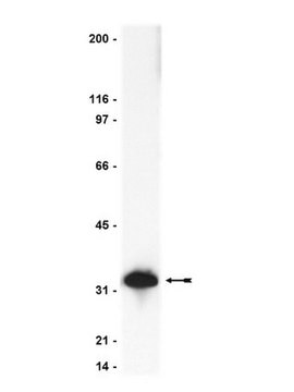
Western Blot Analysis: 5 µg/mL antibody detected p53 on 10 µg of A549 cells treated with UV & TSA lysate.

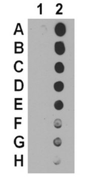
Western Blot (SNAP ID) Analysis: 5 µg/mL antibody detected p53 on 10 µg of recombinant proteins.

Quality

Evaluated by Western Blotting using A549 cells treated with UV & TSA.
Western Blotting Analysis: 5 µg/mL of a representative lot of this antibody detected p53 on 10 µg of A549 cells treated with UV & TSA lysate.

Target description

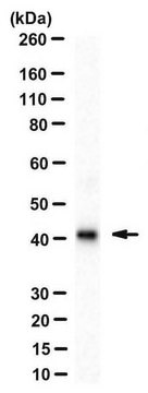
~ 53 kDa observed. Western Blot of A549 cells treated with UV & TSA show bands at ~48 kDa and ~14 kDa which are likely breakdown products of p53 reported for UV treated cells. (Sadji-Ouatas, 2002)

Linkage

Replaces: 06-915

Physical form

Affinity purified
Purified rabbit polyclonal in buffer containing 0.1 M Tris-Glycine (pH 7.4), 150 mM NaCl with 0.05% sodium azide.

Storage and Stability

Stable for 1 year at 2-8°C from date of receipt.

Analysis Note

Control
Recombinant proteins

Other Notes

Concentration: Please refer to the Certificate of Analysis for the lot-specific concentration.

Disclaimer

Unless otherwise stated in our catalog or other company documentation accompanying the product(s), our products are intended for research use only and are not to be used for any other purpose, which includes but is not limited to, unauthorized commercial uses, in vitro diagnostic uses, ex vivo or in vivo therapeutic uses or any type of consumption or application to humans or animals.

Not finding the right product?  

Try our Product Selector Tool.

wgk_germany

WGK 1

flash_point_f

Not applicable

flash_point_c

Not applicable


Certificates of Analysis (COA)

Search for Certificates of Analysis (COA) by entering the products Lot/Batch Number. Lot and Batch Numbers can be found on a product’s label following the words ‘Lot’ or ‘Batch’.

Already Own This Product?

Find documentation for the products that you have recently purchased in the Document Library.

Visit the Document Library

Hyo-Kyoung Choi et al.
Human molecular genetics, 24(4), 1127-1141 (2014-10-12)
Deciphering the molecular basis of neuronal cell death is a central issue in the etiology of neurodegenerative diseases, such as Parkinson's and Alzheimer's. Dysregulation of p53 levels has been implicated in neuronal apoptosis. The role of histone deacetylase 3 (HDAC3)
Shunsheng Zheng et al.
Cancer research, 77(16), 4342-4354 (2017-06-29)
Chemotoxicity due to unwanted p53 activation in the bone marrow remains an unmet clinical challenge. Doxorubicin, a first-line chemotherapy drug, often causes myelosuppression in patients, thus limiting its effectiveness. In this study, we discovered that C646, a reversible p300 inhibitor
Zhi Wen et al.
Molecular and cellular biology, 32(1), 26-35 (2011-10-26)
Since inactivation of tumor suppressor p53 functions is one of the most common features of human cancer cells, restoring p53 expression and activity is an important focus in cancer therapy. Here we report identification of photoreceptor-specific nuclear receptor (PNR)/NR2E3 as
Lu Yang et al.
Communications biology, 4(1), 1373-1373 (2021-12-10)
Tumor suppressor p53, a critical regulator of cell fate, is frequently mutated in cancer. Mutation of p53 abolishes its tumor-suppressing functions or endows oncogenic functions. We recently found that p53 binds via its proline-rich domain to peptidase D (PEPD) and
Stephanie A Luff et al.
PloS one, 13(9), e0203991-e0203991 (2018-09-20)
Megakaryocytes (Mks) derive from hematopoietic stem and progenitor cells (HSPCs) in the bone marrow and develop into large, polyploid cells that eventually give rise to platelets. As Mks mature, they migrate from the bone marrow niche into the vasculature, where

Our team of scientists has experience in all areas of research including Life Science, Material Science, Chemical Synthesis, Chromatography, Analytical and many others.

Contact Technical Service