Skip to Content
MilliporeSigma
All Photos(1)

Documents

06-597

Sigma-Aldrich

Anti-phospho-Histone H1 Antibody

Upstate®, from rabbit

Sign Into View Organizational & Contract Pricing


About This Item

UNSPSC Code:
12352203
eCl@ss:
32160702
NACRES:
NA.41

biological source

rabbit

Quality Level

antibody form

affinity purified immunoglobulin

antibody product type

primary antibodies

clone

polyclonal

species reactivity

bovine, human

packaging

antibody small pack of 25 μg

manufacturer/tradename

Upstate®

technique(s)

immunocytochemistry: suitable
western blot: suitable

isotype

IgG

NCBI accession no.

UniProt accession no.

shipped in

dry ice

target post-translational modification

phosphorylation (not specified)

Gene Information

human ... HIST1H1C(3006)

Related Categories

General description

Histone H1 is one of the five main histone proteins involved in the structure of chromatin in eukaryotic cells. Featuring a main globular domain and a long N-terminal tail, H1 is involved with the structure of the nucleosomes of the ′beads on a string′ structure.

The N-terminal tail of histone H1 protrudes from the globular nucleosome core and can undergo several different types of epigenetic modifications that influence cellular processes. These modifications include the covalent attachment of methyl or acetyl groups to lysine and arginine amino acids and the phosphorylation of serine or threonine.

Specificity

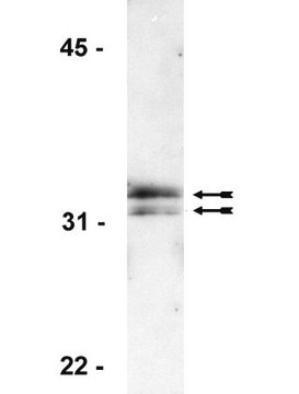
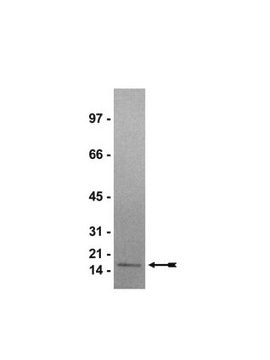
Recognizes phospho-Histone H1, Mr 32 kDa. Additional proteins were also detected, Mr 66 and 100 kDa.

Immunogen

Hyper-phosphorylated isoform of tetrahymena Histone H1 purified by cation-exchange HPLC

Application

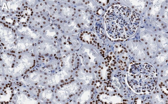
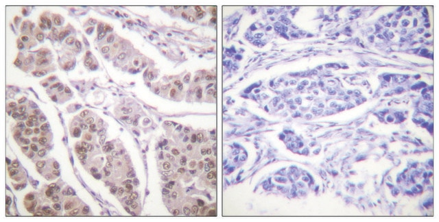
Anti-phospho-Histone H1 Antibody is a high quality Rabbit Polyclonal Antibody for the detection of phospho-Histone H1 & has been validated in ICC & WB.
Research Category
Epigenetics & Nuclear Function
Research Sub Category
Histones

Quality

Immunoblot Analysis: 0.5-2 μg/mL detected phosphorylated Histone H1 from acid extracts of colcemid treated HeLa cells but not untreated HeLa cells (Catalog # 17-306).

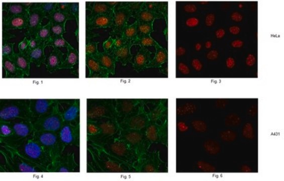
Immunocytochemistry: 5-10 μg/mL showed positive immunostaining for Histone H1 in mitotic HeLa cells fixed with 95% ethanol/5% glacial acetic acid (v/v).

Target description

32 kDa

Physical form

Format: Purified
Immunoaffinity Purified immunoglobulin in 0.02M Phosphate Buffer, 0.25 M NaCl, pH 7.6 with 15 mg/mL BSA containing no preservatives.
Protein A purified

Storage and Stability

Maintain for 2 years at -20°C from date of shipment. Aliquot to avoid repeated freezing and thawing. For maximum recovery of product, centrifuge the original vial after thawing and prior to removing the cap.

Analysis Note

Control
Positive Antigen Control: Catalog #12-309, Hela cell nuclear extract. Add an equal volume of Laemmli reducing sample buffer to 10 μL of extract and boil for 5 minutes to reduce the preparation. Load 20 μg of reduced extract per lane for minigels.

Other Notes

Concentration: Please refer to the Certificate of Analysis for the lot-specific concentration.

Legal Information

UPSTATE is a registered trademark of Merck KGaA, Darmstadt, Germany

Disclaimer

Unless otherwise stated in our catalog or other company documentation accompanying the product(s), our products are intended for research use only and are not to be used for any other purpose, which includes but is not limited to, unauthorized commercial uses, in vitro diagnostic uses, ex vivo or in vivo therapeutic uses or any type of consumption or application to humans or animals.

Still not finding the right product?  

Give our Product Selector Tool a try.


Certificates of Analysis (COA)

Search for Certificates of Analysis (COA) by entering the products Lot/Batch Number. Lot and Batch Numbers can be found on a product’s label following the words ‘Lot’ or ‘Batch’.

Already Own This Product?

Find documentation for the products that you have recently purchased in the Document Library.

Visit the Document Library

Generation and characterization of novel antibodies highly selective for phosphorylated linker histone H1 in Tetrahymena and HeLa cells.
Lu, M J, et al.
Chromosoma, 103, 111-121 (1994)
Diego Piedrahita et al.
Frontiers in cellular neuroscience, 9, 498-498 (2016-01-19)
β-site APP cleaving enzyme 1 (BACE1) initiates APP cleavage, which has been reported to be an inducer of tau pathology by altering proteasome functions in Alzheimer's disease (AD). However, the exact relationship between BACE1 and PHF (Paired Helical Filaments) formation
Steven J T Jackson et al.
Carcinogenesis, 25(2), 219-227 (2003-10-28)
Sulforaphane (SUL), an isothiocyanate found in broccoli and other cruciferous vegetables, has been shown to induce phase II detoxification enzymes, inhibit chemically induced mammary tumors in rats, and more recently to induce cell cycle arrest and apoptosis in cancer cells
Cyclin-dependent kinase 1-mediated Bcl-xL/Bcl-2 phosphorylation acts as a functional link coupling mitotic arrest and apoptosis.
David T Terrano,Meenakshi Upreti,Timothy C Chambers
Molecular and cellular biology null
A simplified formaldehyde fixation and immunoprecipitation technique for studying protein-DNA interactions.
Dedon, P C, et al.
Analytical biochemistry, 197, 83-90 (1991)

Our team of scientists has experience in all areas of research including Life Science, Material Science, Chemical Synthesis, Chromatography, Analytical and many others.

Contact Technical Service