Skip to Content
MilliporeSigma
All Photos(2)

Documents

AB1580-I

Sigma-Aldrich

Anti-Mu Opioid Receptor Antibody

from rabbit, purified by affinity chromatography

Synonym(s):

Mu-type opioid receptor, M-OR-1, MOR-1, Mu opiate receptor, Mu opioid receptor, MOP, hMOP, Anti-Opioid Receptor μ

Sign Into View Organizational & Contract Pricing


About This Item

UNSPSC Code:
12352203
eCl@ss:
32160702
NACRES:
NA.41

biological source

rabbit

Quality Level

antibody form

affinity isolated antibody

antibody product type

primary antibodies

clone

polyclonal

purified by

affinity chromatography

species reactivity

mouse, human

species reactivity (predicted by homology)

bovine (based on 100% sequence homology), porcine (based on 100% sequence homology), rat (based on 100% sequence homology)

technique(s)

immunohistochemistry: suitable (paraffin)
western blot: suitable

NCBI accession no.

UniProt accession no.

shipped in

wet ice

target post-translational modification

unmodified

Gene Information

human ... OPRM1(4988)

General description

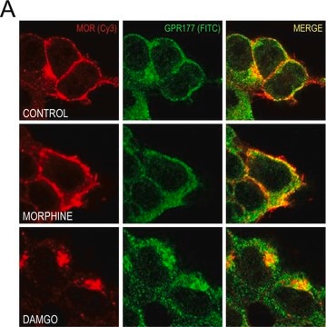
Mu Opioid Receptor (MOP), also called Mu-type opioid receptor (MOR-1), is a receptor for natural and synthetic opioids such as beta-endorphin, endomorphin, morphine, heroin, DAMGO, fentanyl, etorphine, buprenorphin and methadone. MOP functions as a class A G-protein coupled receptor (GPCR), but its down-regulation pathways vary with the agonist and may or may not be independent of G-protein coupling. Endogenous ligands induce rapid desensitization, endocytosis and recycling, as opposed to synthetic ligands, like morphine, that trigger only low desensitization and endocytosis. Hetero-oligomerization with other GPCRs can modulate agonist binding, signaling and trafficking properties. MOP is expressed in the brain and is involved in neurogenesis. MOP is the main physiological target for most clinically important opioid analgesics.

Specificity

This antibody recognizes the C-terminus of Mu Opioid Receptor. This antibody will recognize isoform 1 (45 kDa), isform 10 (55 kDa), isoform 12 (34 kDa), and isoform 13 (36 kDa).

Immunogen

KLH-conjugated linear peptide corresponding to the C-terminus of human Mu Opioid Receptor.

Application

Detect Opioid Receptor using this rabbit polyclonal antibody, Anti-Mu Opioid Receptor Antibody validated for use in western blotting & IHC (Paraffin).
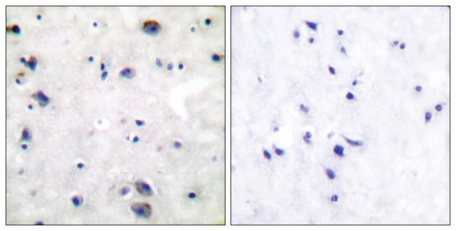
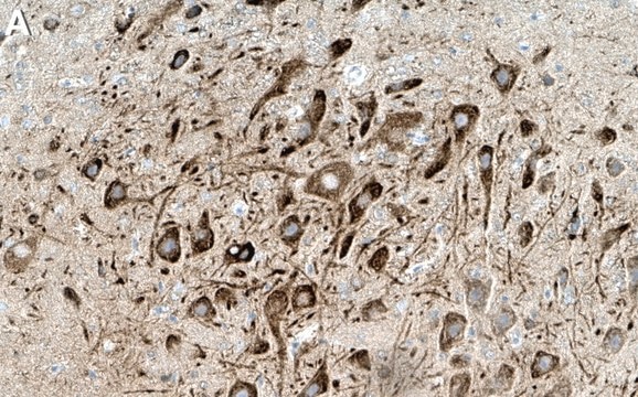
Immunohistochemistry Analysis: A 1:1,000-4,000 dilution from a representative lot detected Mu Opioid Receptor in human cerebellum, mouse hippocampus, mouse midbrain, and rat midbrain tissue.

Quality

Evaluated by Western Blotting in SH-SY5Y cell lysate.

Western Blotting Analysis: 0.2 µg/mL of this antibody detected Mu Opioid Receptor in 10 µg of SH-SY5Y cell lysate.

Target description

~70 kDa observed. The calculated molecular weight is 45 kDa, however Opioid Receptor mu has been shown as a ~70-90 kDa band in western blots (Ferrer-Alcon, M., et al. (2004). Molecular Brain Research. 121(1-2):114-122).

Linkage

Replaces: AB1580

Other Notes

Concentration: Please refer to the Certificate of Analysis for the lot-specific concentration.

Still not finding the right product?  

Give our Product Selector Tool a try.

Storage Class

12 - Non Combustible Liquids

wgk_germany

WGK 1

flash_point_f

Not applicable

flash_point_c

Not applicable


Certificates of Analysis (COA)

Search for Certificates of Analysis (COA) by entering the products Lot/Batch Number. Lot and Batch Numbers can be found on a product’s label following the words ‘Lot’ or ‘Batch’.

Already Own This Product?

Find documentation for the products that you have recently purchased in the Document Library.

Visit the Document Library

Aysegul Gorur et al.
Journal of cellular physiology, 236(11), 7698-7710 (2021-05-27)
The Mu-opioid receptor (MOR) has been implicated in tumorigenesis and metastasis. Methylnaltrexone (MNTX), an antagonist of MOR, has shown to inhibit tumor growth and metastasis in lung cancer cell lines. The effect of MNTX on other cell lines such as
Fei Zhu et al.
Neuron, 99(4), 781-799 (2018-08-07)
Synapses are found in vast numbers in the brain and contain complex proteomes. We developed genetic labeling and imaging methods to examine synaptic proteins in individual excitatory synapses across all regions of the mouse brain. Synapse catalogs were generated from

Our team of scientists has experience in all areas of research including Life Science, Material Science, Chemical Synthesis, Chromatography, Analytical and many others.

Contact Technical Service