Skip to Content
MilliporeSigma
All Photos(4)

Documents

AB9668

Sigma-Aldrich

Anti-Tau phospho Threonine 231 Antibody

Chemicon®, from rabbit

Synonym(s):

Anti-DDPAC, Anti-FTDP-17, Anti-MAPTL, Anti-MSTD, Anti-MTBT1, Anti-MTBT2, Anti-PPND, Anti-PPP1R103, Anti-TAU, Anti-tau-40

Sign Into View Organizational & Contract Pricing


About This Item

UNSPSC Code:
12352203
eCl@ss:
32160702
NACRES:
NA.41

biological source

rabbit

Quality Level

antibody form

affinity purified immunoglobulin

antibody product type

primary antibodies

clone

polyclonal

purified by

affinity chromatography

species reactivity

human

manufacturer/tradename

Chemicon®

technique(s)

western blot: suitable

UniProt accession no.

shipped in

dry ice

target post-translational modification

phosphorylation (pThr231)

Gene Information

human ... MAPT(4137)

General description

Tau is a neuronal microtubule-associated protein found predominantly on axons and functions to promote tubulin polymerization and stabilize microtubules. Tau, in its hyperphosphorylated form, is the major component of paired helical filaments (PHF), the building block of neurofibrillary lesions in Alzheimer′s disease (AD) brain. Hyperphosphorylated Tau is also found in neurofibrillary lesions in a range of other central nervous system disorders. Hyperphosphorylation impairs the microtubule binding function of Tau, resulting in the destabilization of microtubules in AD brains, ultimately leading to the degeneration of the affected neurons. Numerous serine/threonine kinases, including GSK-3beta, protein kinase A (PKA), cyclin-dependent kinase 5 (cdk5) and casein kinase II (CK2), phosphorylate Tau. Threonine 231 is phosphorylated by GSK-3beta, cdk5 and cdk1 and has been shown to be involved in the pre-tangle process of AD.

Specificity

Tau phosphoThreonine 231. The antibody recognizes Tau pThreonine 231 in samples of recombinant human Tau treated with GSK-3beta for 45 minutes. The reactivity of the antibody is blocked with the pThreonine 231 peptide but not the non-phosphopeptide or a generic phosphoThreonine-containing peptide.

Immunogen

Synthetic peptide of amino acids surrounding the phosphoThreonine 231 site of human Tau.

Application

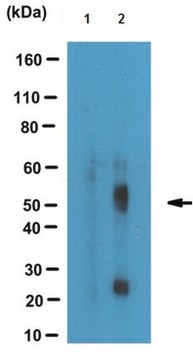
Anti-Tau phospho Threonine 231 Antibody detects level of Tau phospho Threonine 231 & has been published & validated for use in WB.
Research Category
Neuroscience
Research Sub Category
Neurodegenerative Diseases
Western blot: 1:1,000. Suggested blocking buffer is 5% BSA-TBST for one hour at room temperature. Suggested antibody dilution buffer is 1% BSA-TBST. Suggested antibody incubation time is 2 hours at room temperature..

Optimal working dilutions must be determined by the end user.

Physical form

Affinity purified immunoglobulin. Liquid in Dulbecco′s PBS (without Mg2+ and Ca2+), pH 7.3, 50% glycerol with 1.0 mg/mL BSA and 0.05% sodium azide.

Storage and Stability

Maintain at -20°C in undiluted for up to 6 months after date of receipt. Avoid repeated freeze/thaw cycles. Do not store in a self defrosting freezer.

Legal Information

CHEMICON is a registered trademark of Merck KGaA, Darmstadt, Germany

Disclaimer

Unless otherwise stated in our catalog or other company documentation accompanying the product(s), our products are intended for research use only and are not to be used for any other purpose, which includes but is not limited to, unauthorized commercial uses, in vitro diagnostic uses, ex vivo or in vivo therapeutic uses or any type of consumption or application to humans or animals.

Not finding the right product?  

Try our Product Selector Tool.

wgk_germany

WGK 2


Certificates of Analysis (COA)

Search for Certificates of Analysis (COA) by entering the products Lot/Batch Number. Lot and Batch Numbers can be found on a product’s label following the words ‘Lot’ or ‘Batch’.

Already Own This Product?

Find documentation for the products that you have recently purchased in the Document Library.

Visit the Document Library

Increased Tau Phosphorylation and Impaired Brain Insulin/IGF Signaling in Mice Fed a High Fat/High Cholesterol Diet.
Bhat, Narayan R and Thirumangalakudi, Lakshmi
Journal of Alzheimer'S Disease (2013)
Fei Liu et al.
The Journal of biological chemistry, 280(3), 1790-1796 (2004-11-18)
Protein phosphatase (PP) 5 is highly expressed in the mammalian brain, but few physiological substrates have yet been identified. Here, we investigated the kinetics of dephosphoryation of phospho-tau by PP5 and found that PP5 had a K(m) of 8-13 microm
I Tesseur et al.
The American journal of pathology, 156(3), 951-964 (2000-03-07)
Epidemiological studies have established that the epsilon 4 allele of the ApoE gene (ApoE4) constitutes an important risk factor for Alzheimer's disease and might influence the outcome of central nervous system injury. The mechanism by which ApoE4 contributes to the
Abhay P Sagare et al.
Nature communications, 4, 2932-2932 (2013-12-18)
Pericytes are cells in the blood-brain barrier that degenerate in Alzheimer's disease (AD), a neurological disorder associated with neurovascular dysfunction, abnormal elevation of amyloid β-peptide (Aβ), tau pathology and neuronal loss. Whether pericyte degeneration can influence AD-like neurodegeneration and contribute
C X Gong et al.
The Journal of biological chemistry, 275(8), 5535-5544 (2000-02-22)
Hyperphosphorylated tau, which is the major protein of the neurofibrillary tangles in Alzheimer's disease brain, is most probably the result of an imbalance of tau kinase and phosphatase activities in the affected neurons. By using metabolically competent rat brain slices

Our team of scientists has experience in all areas of research including Life Science, Material Science, Chemical Synthesis, Chromatography, Analytical and many others.

Contact Technical Service