Skip to Content
MilliporeSigma
All Photos(2)

Documents

ABN1698

Sigma-Aldrich

Anti-Aspa/Nur7 Antibody

from rabbit, purified by affinity chromatography

Synonym(s):

Aspartoacylase, Aminoacylase-2, ACY-2, Aspa/Nur7, Anti-Aspartoacylase

Sign Into View Organizational & Contract Pricing


About This Item

UNSPSC Code:
12352203
eCl@ss:
32160702
NACRES:
NA.41

biological source

rabbit

Quality Level

antibody form

purified antibody

antibody product type

primary antibodies

clone

polyclonal

purified by

affinity chromatography

species reactivity

human, mouse, rat

technique(s)

immunohistochemistry: suitable
western blot: suitable

NCBI accession no.

UniProt accession no.

shipped in

wet ice

target post-translational modification

unmodified

Gene Information

human ... ASPA(443)

Related Categories

General description

Aspartoacylase (EC 3.5.1.15; UniProt Q8R3P0; also known as Acy-2, Aminoacylase-2, Nur-7) is encoded by the Aspa (also known as Acy2) gene (Gene ID 11484) in murine species. Aspartoacylase catalyzes the deacetylation of N-acetylaspartate (NAA) to generate free acetate in the central nervous system (CNS). Mutations in the ASPA gene cause the autosomal recessive neurodegenerative Canavan disease (CD) that results in inadequate lipid/myelin synthesis during development. Immunohistochemical staining reveals that aspartoacylase colocalizes with the oligodendrocyte marker CC1 throughout the brain, indicating its role in myelination.

Immunogen

N-terminally his-tagged full-length recombinant mouse Aspa/Nur7.

Application

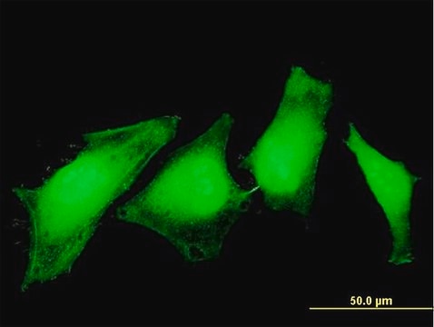
Immunohistochemistry Analysis: A representative lot detected Aspa/Nur7-positive oligodendrocytes in corpus callosum/external capsule using free-floating mouse brain sections (Courtesy of Dr. John R. Moffett, Uniformed Services University of the Health Sciences).
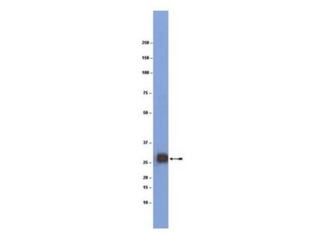
Western Blotting Analysis: A representative lot detected Aspa/Nur7 in the cytosol, but not myelin, fraction from rat brain tissue homogenate (Madhavarao, C.N., et al. (2004). J Comp Neurol. 472(3):318-329).
Immunohistochemistry Analysis: A representative lot detected Aspa/Nur7 expression pattern in various regions of rat forebrain, including corpus callosum, cerebral cortex, hippocampal commissure (hc), fimbria, and anterior commissure (Madhavarao, C.N., et al. (2004). J Comp Neurol. 472(3):318-329).
Immunohistochemistry Analysis: A representative lot detected similar Aspa/Nur7 expression pattern as CC1 in various regions of rat forebrain, including the Purkinjie & axonal fiber layers of cerebellum, corpus callosum, as well as layer 2 of primary somatosensory cortex (Madhavarao, C.N., et al. (2004). J Comp Neurol. 472(3):318-329).
Research Category
Neuroscience
Research Sub Category
Developmental Signaling
This Anti-Aspa/Nur7 Antibody is validated for use in Western Blotting, Immunohistochemistry for the detection of Aspa/Nur7.

Quality

Evaluated by Western Blotting in rat brain tissue lysate.

Western Blotting Analysis: 1.0 µg/mL of this antibody detected Aspa/Nur7 in 10 µg of rat brain tissue lysate.

Target description

~37 kDa observed

Physical form

Protein G Purified
Purified rabbit polyclonal antibody in buffer containing 0.1 M Tris-Glycine (pH 7.4), 150 mM NaCl with 0.05% sodium azide.

Storage and Stability

Stable for 1 year at 2-8°C from date of receipt.

Other Notes

Concentration: Please refer to lot specific datasheet.

Disclaimer

Unless otherwise stated in our catalog or other company documentation accompanying the product(s), our products are intended for research use only and are not to be used for any other purpose, which includes but is not limited to, unauthorized commercial uses, in vitro diagnostic uses, ex vivo or in vivo therapeutic uses or any type of consumption or application to humans or animals.

Not finding the right product?  

Try our Product Selector Tool.

wgk_germany

WGK 1

flash_point_f

Not applicable

flash_point_c

Not applicable


Certificates of Analysis (COA)

Search for Certificates of Analysis (COA) by entering the products Lot/Batch Number. Lot and Batch Numbers can be found on a product’s label following the words ‘Lot’ or ‘Batch’.

Already Own This Product?

Find documentation for the products that you have recently purchased in the Document Library.

Visit the Document Library

Immunohistochemical localization of aspartoacylase in the rat central nervous system.
Madhavarao, CN; Moffett, JR; Moore, RA; Viola, RE; Namboodiri, MA; Jacobowitz, DM
The Journal of Comparative Neurology null
Simon Pan et al.
Nature neuroscience, 23(4), 487-499 (2020-02-12)
Experience-dependent myelination is hypothesized to shape neural circuit function and subsequent behavioral output. Using a contextual fear memory task in mice, we demonstrate that fear learning induces oligodendrocyte precursor cells to proliferate and differentiate into myelinating oligodendrocytes in the medial
Chih-Fen Hu et al.
Frontiers in immunology, 12, 638381-638381 (2021-04-20)
While oxidative stress has been linked to multiple sclerosis (MS), the role of superoxide-producing phagocyte NADPH oxidase (Nox2) in central nervous system (CNS) pathogenesis remains unclear. This study investigates the impact of Nox2 gene ablation on pro- and anti-inflammatory cytokine
Anthony Fernández-Castañeda et al.
Cell, 185(14), 2452-2468 (2022-06-30)
COVID survivors frequently experience lingering neurological symptoms that resemble cancer-therapy-related cognitive impairment, a syndrome for which white matter microglial reactivity and consequent neural dysregulation is central. Here, we explored the neurobiological effects of respiratory SARS-CoV-2 infection and found white-matter-selective microglial
Zhixin Lei et al.
The Journal of neuroscience : the official journal of the Society for Neuroscience, 40(33), 6444-6456 (2020-07-15)
Previous studies demonstrate that activation of pancreatic ER kinase (PERK) protects oligodendrocytes against inflammation in the experimental autoimmune encephalomyelitis (EAE) model of multiple sclerosis (MS). Interestingly, data indicate that the cytoprotective effects of PERK activation on oligodendrocytes during EAE are

Our team of scientists has experience in all areas of research including Life Science, Material Science, Chemical Synthesis, Chromatography, Analytical and many others.

Contact Technical Service