Skip to Content
MilliporeSigma
All Photos(2)

Documents

ABS1616

Sigma-Aldrich

Anti-CGI-58 Antibody

serum, from rabbit

Synonym(s):

1-acylglycerol-3-phosphate O-acyltransferase ABHD5 (EC:2.3.1.51), Abhydrolase domain-containing protein 5, Lipid droplet-binding protein CGI-58, Protein CGI-58, CGI-58

Sign Into View Organizational & Contract Pricing


About This Item

UNSPSC Code:
12352203
eCl@ss:
32160702
NACRES:
NA.41

biological source

rabbit

Quality Level

antibody form

serum

antibody product type

primary antibodies

clone

polyclonal

species reactivity

mouse

technique(s)

immunocytochemistry: suitable
western blot: suitable

NCBI accession no.

UniProt accession no.

shipped in

dry ice

target post-translational modification

unmodified

Gene Information

mouse ... Abhd5(67469)

General description

1-acylglycerol-3-phosphate O-acyltransferase ABHD5 (EC:2.3.1.51; UniProt Q9DBL9; also known as Abhydrolase domain-containing protein 5, CGI-58, Comparative gene identification-58, Lipid droplet-binding protein CGI-58) is encoded by the Abhd5 (also known as CDS, IECN5, NCIE2, 1300003D03Rik, 2010002J10Rik) gene in murine species (Gene ID 67469). The comparative gene identification-58 (CGI-58) functions as a cofactor of adipose triglyceride lipase/ATGL-mediated triacylglycerol (TG) hydrolytic activity, which is essential for efficient catabolism of cellular TG stores. Mutations in the genes coding for ATGL or CGI-58 in humans cause neutral lipid storage disease characterized by TG accumulation in multiple tissues. CGI-58 gene mutations provoke severe ichthyosis and hepatosteatosis in humans and mice. Mice lacking CGI-58 exclusively in muscle (CGI-58KOM) show TG accumulation in both skeletal and cardiac muscle tissues and develop severe cardiac steatosis and cardiomyopathy as a result of impaired TG catabolism and mitochondrial fatty acid oxidation. Immunocytochemistry analysis reveals diffuse cytoplasmic staining of CGI-58 in 3T3-L1 preadipocytes, while CGI-58 staining is found exclusively on the surface of lipid droplets in differentiated 3T3-L1 adipocytes. CGI-58 binds to lipid droplets by interacting with perilipin C-terminal sequence, while activation of cellular PKA disperses CGI-58 localization from the lipid droplets to cytosol.

Immunogen

His-tagged recombinant protein corresponding to mouse CGI-58.

Application

Detect CGI-58 using this Anti-CGI-58 Antibody validated for use in Western Blotting and Immunocytochemistry.
Immunocytochemistry Analysis: A 1:5,000 dilution from a representative lot detected CGI-58 in 3T3-L1 adipocytes (Data courtesy of Dawn Brasaemle, Rutgers University).
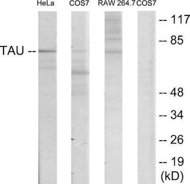
Western Blotting Analysis: A representative lot detected recombinant CGI-58, as well as endogenous CGI-58 in differentiated 3T3-L1 adipocytes, and 3T3-L1 preadipocytes stably expressing ectopic perilipin A or perilipin B (Subramanian, V., et al. (2004). J. Biol. Chem. 279(40):42062-42071).
Immunocytochemistry Analysis: A representative lot detected CGI-58 in 3T3-L1 adipocytes, 3T3-L1 preadipocytes stably expressing perilipin A (Subramanian, V., et al. (2004). J. Biol. Chem. 279:42062-42071).
Western Blotting Analysis: A representative lot detected CGI-58 in mouse cardiac muscle, liver, white and brown adipose tissue (Zierler, K.A., et al. (2013). J. Biol. Chem. 288(14):9892-9904).
Western Blotting Analysis: A representative lot detected CGI-58 in in mouse OP9 adipocytes and brown adipose tissue (Skinner, J.R., et al. (2013). Adipocyte. 2(2):80-86).
Research Category
Signaling
Research Sub Category
Signaling Neuroscience

Quality

Evaluated by Western Blotting in differentiated 3T3-L1 adipocytes cell lysate.

Western Blotting Analysis: A 1:1,000 of this antibody detected CGI-58 in 10 µg of differentiated 3T3-L1 adipocytes cell lysate.

Target description

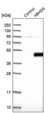
~42 kDa observed. CGI-58 migrates with an apparent molecular size slightly larger than the calculated molecular weight of 39.2 kDa for mouse CGI-58.

Physical form

Rabbit polyclonal serum with 0.05% sodium azide.
Unpurified

Storage and Stability

Stable for 1 year at -20°C from date of receipt.
Handling Recommendations: Upon receipt and prior to removing the cap, centrifuge the vial and gently mix the solution. Aliquot into microcentrifuge tubes and store at -20°C. Avoid repeated freeze/thaw cycles, which may damage IgG and affect product performance.

Other Notes

Concentration: Please refer to lot specific datasheet.

Disclaimer

Unless otherwise stated in our catalog or other company documentation accompanying the product(s), our products are intended for research use only and are not to be used for any other purpose, which includes but is not limited to, unauthorized commercial uses, in vitro diagnostic uses, ex vivo or in vivo therapeutic uses or any type of consumption or application to humans or animals.

Not finding the right product?  

Try our Product Selector Tool.

wgk_germany

WGK 1


Certificates of Analysis (COA)

Search for Certificates of Analysis (COA) by entering the products Lot/Batch Number. Lot and Batch Numbers can be found on a product’s label following the words ‘Lot’ or ‘Batch’.

Already Own This Product?

Find documentation for the products that you have recently purchased in the Document Library.

Visit the Document Library

Boubacar Benziane et al.
Journal of lipid research, 58(12), 2324-2333 (2017-10-27)
Diacylglycerol kinases (DGKs) regulate the balance between diacylglycerol (DAG) and phosphatidic acid. DGKζ is highly abundant in skeletal muscle and induces fiber hypertrophy. We hypothesized that DGKζ influences functional and metabolic adaptations in skeletal muscle and whole-body fuel utilization. DAG

Our team of scientists has experience in all areas of research including Life Science, Material Science, Chemical Synthesis, Chromatography, Analytical and many others.

Contact Technical Service