Skip to Content
MilliporeSigma
All Photos(3)

Documents

MAB1959

Sigma-Aldrich

Anti-Integrin β1 Antibody, clone P5D2

clone P5D2, Chemicon®, from mouse

Synonym(s):

CD29, MAB1959Z

Sign Into View Organizational & Contract Pricing


About This Item

UNSPSC Code:
12352203
eCl@ss:
32160702
NACRES:
NA.41

biological source

mouse

Quality Level

antibody form

purified immunoglobulin

antibody product type

primary antibodies

clone

P5D2, monoclonal

species reactivity

human

manufacturer/tradename

Chemicon®

technique(s)

ELISA: suitable
flow cytometry: suitable
immunocytochemistry: suitable
immunohistochemistry: suitable
immunoprecipitation (IP): suitable

isotype

IgG2bκ

UniProt accession no.

shipped in

wet ice

target post-translational modification

unmodified

Gene Information

human ... ITGB1(3688)

Related Categories

General description

Integrin beta 1, also known as CD29, is a 130 kDa transmembrane glycoprotein that forms noncovalent complexes with various Integrin alpha subunits (including alpha 1, alpha 2, alpha 3, alpha 4, alpha 5, and alpha 6) to form the functional receptors that bind to specific extracellular matrix proteins. Integrin receptors are involved in the regulation of a variety of important biological functions, including embryonic development, wound repair, hemostasis, and prevention of programmed cell death. They are also implicated in abnormal pathological states such as tumor directed angiogenesis, tumor cell growth, and metastasis. These heterodimeric receptors bridge the cytoplasmic actin cytoskeleton with proteins present in the extracellular matrix and/or on adjacent cells. Interactions between integrins and the extracellular matrix lead to activation of signal transduction pathways and regulation of gene expression.

Specificity

Reacts with Human Integrin Beta 1.

Immunogen

Human keratinocytes derived from skin.

Application

Immunoprecipitation: A representative lot of this antibody clone was used in immunoprecipitation.

Immunohistochemistry: A representative lot of this antibody clone was used in immunohistochemistry (acetone fixation, no paraffin embedding).

ELISA: A representative lot of this antibody was used in ELISA.

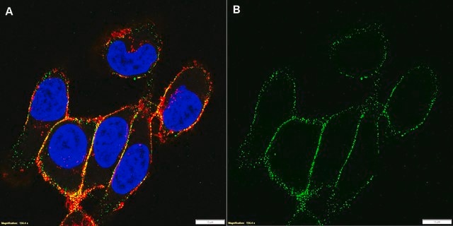
Immunocytochemistry: A representative lot of this antibody clone was used in immunocytochemistry (paraformaldehyde fixation at less than 4%).

Functional Activity Assay: A representative lot of this antibody clone was used in cell attachment assay of SV-HFO cells with a characteristic spread morphology. In the presence of function-blocking mAbs to β1 integrin (P5D2), the cells attached but no longer spread, and displayed a rounded morphology with many cytoplasmic projections (Iba, K. et al., 2000).
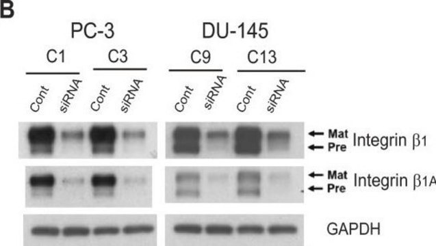
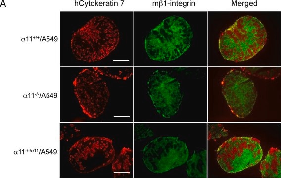
Anti-Integrin β1 Antibody, clone P5D2 detects level of Integrin β1 & has been published & validated for use in ELISA, FC, IC, IH & IP.
Research Category
Cell Structure
Research Sub Category
Integrins

Quality

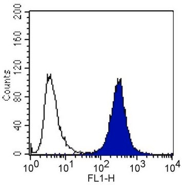
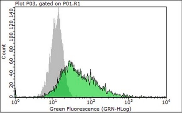
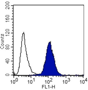
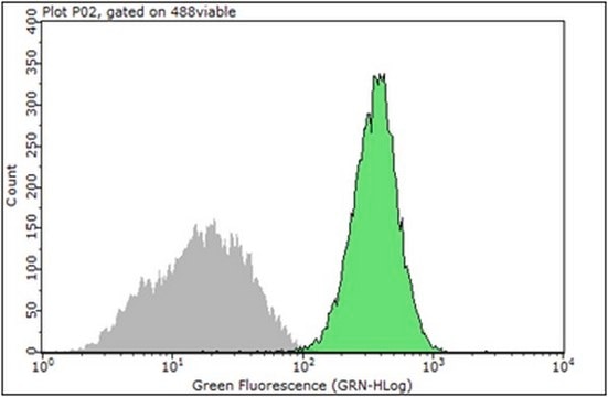
Flow Cytometry Analysis:
4 µg of the antibody was used to detect Integrin β1 in 1x10^6 A431 cells.
4 µg of the antibody was used to detect Integrin β1 in 1x10^6 HeLa cells.

Physical form

Format: Purified
Protein A purified
Purified immunoglobulin in 0.02M PB, 0.25M NaCl, pH 7.6, 0.1% sodium azide.

Storage and Stability

Maintain for 1 year at 2–8°C from date of shipment. Aliquot to avoid repeated freezing and thawing. For maximum recovery of product, centrifuge the original vial after thawing and prior to removing the cap.

Analysis Note

Control
Human tonsil, human skin tissue

A431 & HeLa Cells

Other Notes

Concentration: Please refer to the Certificate of Analysis for the lot-specific concentration.

Legal Information

CHEMICON is a registered trademark of Merck KGaA, Darmstadt, Germany

Disclaimer

Unless otherwise stated in our catalog or other company documentation accompanying the product(s), our products are intended for research use only and are not to be used for any other purpose, which includes but is not limited to, unauthorized commercial uses, in vitro diagnostic uses, ex vivo or in vivo therapeutic uses or any type of consumption or application to humans or animals.

Still not finding the right product?  

Give our Product Selector Tool a try.

Storage Class

10 - Combustible liquids

wgk_germany

WGK 2

flash_point_f

Not applicable

flash_point_c

Not applicable


Certificates of Analysis (COA)

Search for Certificates of Analysis (COA) by entering the products Lot/Batch Number. Lot and Batch Numbers can be found on a product’s label following the words ‘Lot’ or ‘Batch’.

Already Own This Product?

Find documentation for the products that you have recently purchased in the Document Library.

Visit the Document Library

S Gronthos et al.
Proceedings of the National Academy of Sciences of the United States of America, 97(25), 13625-13630 (2000-11-23)
Dentinal repair in the postnatal organism occurs through the activity of specialized cells, odontoblasts, that are thought to be maintained by an as yet undefined precursor population associated with pulp tissue. In this study, we isolated a clonogenic, rapidly proliferative
Sphingosine-1-phosphate induces a PDGFR-dependent cell detachment via inhibiting beta1 integrin in HEK293 cells.
Alexander Zaslavsky, Song Li, Yan Xu
Febs Letters null
Saubhik Sengupta et al.
FASEB journal : official publication of the Federation of American Societies for Experimental Biology, 17(11), 1570-1572 (2003-06-26)
We have reported previously that levels of lysophosphatidic acid (LPA) are elevated in the blood and ascites from patients with ovarian cancer. LPA stimulates proliferation of ovarian cancer cells and has been proposed as an autocrine growth factor. Here, we
Elizabeth A Waterman et al.
Cancer research, 67(9), 4264-4270 (2007-05-08)
Laminin-332 (formerly laminin-5) and collagen VII are basement membrane proteins expressed at the invasive front of human squamous cell carcinoma (SCC) tumors. These proteins have protumorigenic properties, but whether laminin-332 and collagen VII promote SCC tumors by providing adhesion or
B Baudoux et al.
European journal of cell biology, 79(1), 41-51 (2000-03-11)
Integrins are involved in several ways in keratinocyte physiology, including cell motility. CD9 is a member of the tetraspanin protein family which is found in association with other transmembrane proteins like the integrins. CD9 is expressed in the epidermal tissue

Our team of scientists has experience in all areas of research including Life Science, Material Science, Chemical Synthesis, Chromatography, Analytical and many others.

Contact Technical Service