Skip to Content
MilliporeSigma
All Photos(1)

Documents

MAB4197

Sigma-Aldrich

Anti-Topoisomerase II Antibody, clone KiS1

clone KiS1, Chemicon®, from mouse

Synonym(s):

Ki-S1

Sign Into View Organizational & Contract Pricing


About This Item

UNSPSC Code:
12352203
eCl@ss:
32160702
NACRES:
NA.41

biological source

mouse

Quality Level

antibody form

purified immunoglobulin

clone

KiS1, monoclonal

species reactivity

human

manufacturer/tradename

Chemicon®

technique(s)

flow cytometry: suitable
immunocytochemistry: suitable
immunohistochemistry (formalin-fixed, paraffin-embedded sections): suitable
western blot: suitable

isotype

IgG2a

NCBI accession no.

UniProt accession no.

shipped in

wet ice

target post-translational modification

unmodified

Gene Information

human ... TOP2A(7153)

General description

Topoisomerase II β (M.W 180 kDa) plays important roles in synthesis and transcription of DNA as well as chromosomal segregation during mitosis. It is present ubiquitously in normal cells and is upregulated in tumors and proliferating cells. Topoisomerase II β is also implicated in drug resistance of tumor cells.

Specificity

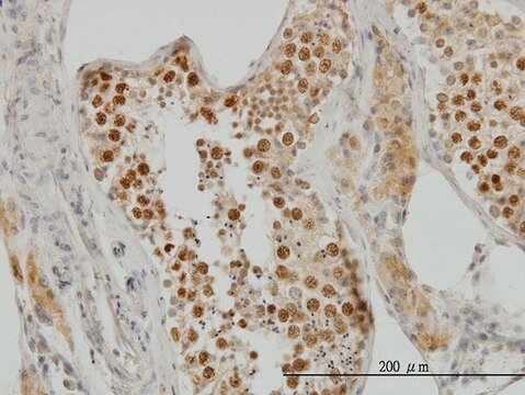
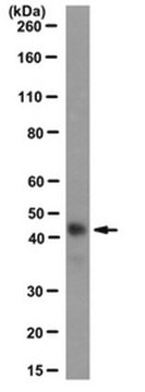
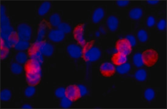
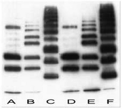
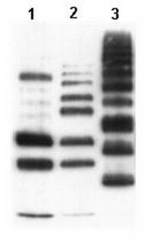
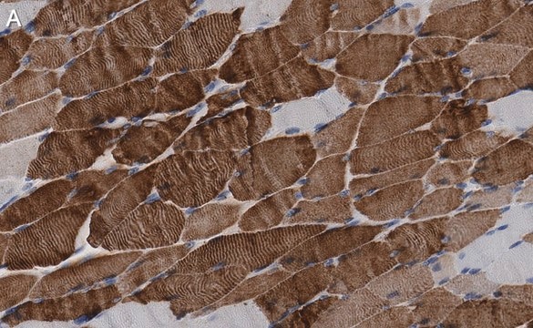
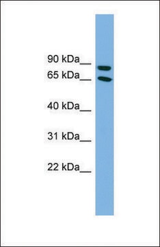
In Immunoprecipitation and Western blot experiments the Ki-S1 antibody recognizes a major protein of 170 kD which was identified as the a isoform of topoisomerase II (Boege et al., 1995; Kreipe et al., 1993). In addition, it has been shown that the Ki-S1 antibody recognizes a carboxyterminal a-isoenzyme specific epitope missing in topoisomerase IIb (Boege et al., 1995). In immunohistochemistry the Ki-S1 antibody shows strong nuclear staining only in proliferating cells. The epitope recognized by the antibody is also detectable in paraffin-embedded tissue sections (Kreipe et al., 1993). Accordingly, it has been shown that the expression of topoisomerase IIa is strongly restricted to proliferating cells (Tsutsui et al., 1993). The topoisomerase IIa antigen is preferentially expressed during G 1 , S, G 2 and M phase of the cell cycle, while resting, non-cycling cells (G 0 phase) lack topoisomerase IIa. In addition, constantly proliferating cells (e.g. cell lines) react positively to topoisomerase IIa during the entire cell-cycle. This specificity of Ki-S1 antibody for proliferating cells might make it a useful tool for determination of the proliferative fraction in solid tumors such as mammary carcinomas (Kreipe et al., 1993; Sampson et al., 1992; Camplejohn et al., 1993; Kreipe et al., 1993; Rasbridge et al., 1994) and gangliomas (Wolf et al., 1994).

Application

Anti-Topoisomerase II Antibody, clone KiS1 is a Mouse Monoclonal Antibody for detection of Topoisomerase II also known as Ki-S1 & has been validated in FC, WB, ICC, IHC, IHC(P).
Western blot: 1-10μg/ml Immunoprecipitaion

Immunocytochemistry: 5-10 μg/ml Immunohistochemistry: 5-10 μg/ml

Flow cytometry

Optimal working dilutions must be determined by end user.

Linkage

Replaces: MABE519

Physical form

Format: Purified

Storage and Stability

Maintain at 2-8°C in undiluted aliquots for up to 6 months.Avoid repeat freeze/thaw cycles.

Other Notes

Concentration: Please refer to the Certificate of Analysis for the lot-specific concentration.

Legal Information

CHEMICON is a registered trademark of Merck KGaA, Darmstadt, Germany

Storage Class

12 - Non Combustible Liquids

wgk_germany

WGK 2

flash_point_f

Not applicable

flash_point_c

Not applicable


Certificates of Analysis (COA)

Search for Certificates of Analysis (COA) by entering the products Lot/Batch Number. Lot and Batch Numbers can be found on a product’s label following the words ‘Lot’ or ‘Batch’.

Already Own This Product?

Find documentation for the products that you have recently purchased in the Document Library.

Visit the Document Library

K Tsutsui et al.
The Journal of biological chemistry, 268(25), 19076-19083 (1993-09-05)
cDNA segments for DNA topoisomerase II were amplified from rat brain RNA after reverse transcription by the polymerase chain reaction, using degenerate oligonucleotide primers deduced from the conserved regions of topoisomerase II of higher eukaryotes. The cDNA product from a
The effects of chemotherapy on morphology, cellular proliferation, apoptosis and oncoprotein expression in primary breast carcinoma.
Rasbridge, S A, et al.
British Journal of Cancer, 70, 335-341 (1994)
N I Valkov et al.
British journal of haematology, 108(2), 331-345 (2000-02-26)
The resistance of several leukaemic and myeloma cell lines (CCRF, L1210, HL-60, KG-1a and RPMI 8226) to VP-16 was found to increase with cell density and to be maximal (3.5- to 39-fold) in plateau phase cell cultures, as measured by
Ganglioglioma: a detailed histopathological and immunohistochemical analysis of 61 cases.
Wolf, H K, et al.
Acta neuropathologica, 88, 166-173 (1994)
KiS1--a novel monoclonal antibody which recognizes proliferating cells: evaluation of its relationship to prognosis in mammary carcinoma.
Sampson, S A, et al.
The Journal of Pathology, 168, 179-185 (1992)

Our team of scientists has experience in all areas of research including Life Science, Material Science, Chemical Synthesis, Chromatography, Analytical and many others.

Contact Technical Service