Skip to Content
MilliporeSigma
All Photos(2)

Documents

MABC1638

Sigma-Aldrich

Anti-AIFM2 Antibody, clone 6D8-11

Sign Into View Organizational & Contract Pricing


About This Item

UNSPSC Code:
12352203
NACRES:
NA.41

biological source

rat

Quality Level

conjugate

unconjugated

antibody form

purified antibody

antibody product type

primary antibodies

clone

6D8, monoclonal

mol wt

calculated mol wt 40.53 kDa
observed mol wt ~40 kDa

purified by

using protein G

species reactivity

human

packaging

antibody small pack of 100 μL

technique(s)

ELISA: suitable
immunocytochemistry: suitable
immunofluorescence: suitable
western blot: suitable

isotype

IgG2a

epitope sequence

Unknown

Protein ID accession no.

UniProt accession no.

shipped in

2-8°C

target post-translational modification

unmodified

Gene Information

General description

Ferroptosis suppressor protein 1 (UniProt: Q9BRQ8; also known as FSP1, Apoptosis-inducing factor homologous mitochondrion-associated inducer of death, AMID, p53-responsive gene 3 protein, AIFM2) is encoded by the AIFM2 (also known as AMID, PRG3) gene (Gene ID: 84883) in human. AIFM2 is a lipid-anchor membrane protein of the FAD-dependent oxidoreductase family that is also detected in the nucleus and mitochondrial membrane. It is highly expressed in heart and moderately in liver and skeletal muscle. Low expression levels are observed in placenta, lung, kidney, and pancreas. It is reported to be down-regulated in a number of human tumors. It serves as a NAD(P)H-dependent oxidoreductase and is involved in cellular oxidative stress response. At the plasma membrane, it catalyzes reduction of coenzyme Q/ubiquinone-10 to ubiquinol-10, a lipophilic radical-trapping antioxidant that prevents lipid oxidative damage and consequently ferroptosis. It is shown to cooperates with GPX4 to suppress phospholipid peroxidation and ferroptosis and its anti-ferroptotic function is reported to be independent of cellular glutathione levels. It also plays a role in mitochondrial stress signaling. Upon induction of oxidative stress, it associates with the lipid peroxidation end product 4-hydroxy-2-nonenal (HNE) forming a lipid adduct. This HNE adduct inactivates the NADH oxidoreductase activity of AIFM2 and facilitates its translocation from mitochondria into the nucleus triggering DNA damage and cell death. (Ref.: Doll, S., et al. (2019). Nature 575(7784); 693-698; Miriyala, S., et al. (2016). Free Radic. Biol. Med. 91; 68-80).

Specificity

Clone 6D8 is a rat monoclonal antibody that detects human Ferroptosis suppressor protein 1 (AIFM2).

Immunogen

SUMO-His-tagged full-length recombinant human Ferroptosis suppressor protein 1 (FSP1/AIFM2).

Application

Quality Control Testing

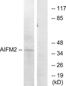
Evaluated by Western Blotting in A549 cell lysate.

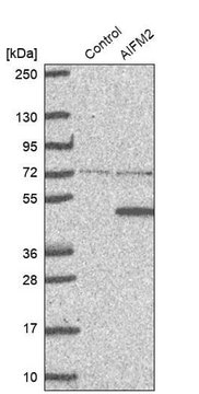
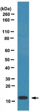
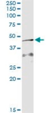
Western Blotting Analysis: A 1:500 dilution of this antibody detected AIFM2 in A549 cell lysate.

Tested Appliacations

Western Blotting Analysis: A 1:500 dilution from a representative lot detected AIFM2 in HepG2 cell lysate.

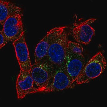
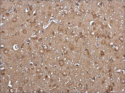
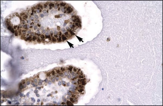
Immunocytochemistry Analysis: A 1:25 Dilution from a representative lot detected AIFM2 in HepG2 cells.

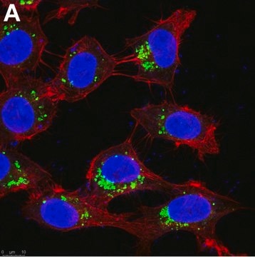
Immunofluorescence Analysis: A representative lot detected AIFM2 in Immunofluorescence applications (Doll, S. et al. (2019). Nature.;575(7784):693-698).

Western Blotting Analysis: A representative lot detected AIFM2 in Western Blotting applications (Doll, S. et al. (2019). Nature.;575(7784):693-698).

ELISA Analysis: A representative lot detected AIFM2 in ELISA applications (Doll, S. et al. (2019). Nature.;575(7784):693-698).

Note: Actual optimal working dilutions must be determined by end user as specimens, and experimental conditions may vary with the end user
Anti-AIFM2, clone 6D8-11, Cat. No. MABC1638, is a rat monoclonal antibody that detects Ferroptosis suppressor protein 1 and is tested for use in ELISA, Immunocytochemistry, Immunofluorescence, and Western Blotting.

Physical form

Purified rat monoclonal antibody IgG2a in buffer containing 0.1 M Tris-Glycine (pH 7.4), 150 mM NaCl with 0.05% sodium azide.

Storage and Stability

Recommend storage at +2°C to +8°C. For long term storage antibodies can be kept at -20°C. Avoid repeated freeze-thaws.

Other Notes

Concentration: Please refer to the Certificate of Analysis for the lot-specific concentration.

Disclaimer

Unless otherwise stated in our catalog or other company documentation accompanying the product(s), our products are intended for research use only and are not to be used for any other purpose, which includes but is not limited to, unauthorized commercial uses, in vitro diagnostic uses, ex vivo or in vivo therapeutic uses or any type of consumption or application to humans or animals.

Not finding the right product?  

Try our Product Selector Tool.

wgk_germany

WGK 1

flash_point_f

Not applicable

flash_point_c

Not applicable


Certificates of Analysis (COA)

Search for Certificates of Analysis (COA) by entering the products Lot/Batch Number. Lot and Batch Numbers can be found on a product’s label following the words ‘Lot’ or ‘Batch’.

Already Own This Product?

Find documentation for the products that you have recently purchased in the Document Library.

Visit the Document Library

Our team of scientists has experience in all areas of research including Life Science, Material Science, Chemical Synthesis, Chromatography, Analytical and many others.

Contact Technical Service