MABE1016
Anti-pan-ADP-ribose binding reagent
from Escherichia coli
Synonym(s):
pan-ADP-ribose binding reagent
Sign Into View Organizational & Contract Pricing
All Photos(3)
About This Item
UNSPSC Code:
12352203
eCl@ss:
32160405
NACRES:
NA.41
Recommended Products
biological source
Escherichia coli
Quality Level
antibody form
purified antibody
antibody product type
primary antibodies
species reactivity
human, mouse
species reactivity (predicted by homology)
all
technique(s)
dot blot: suitable
immunoprecipitation (IP): suitable
western blot: suitable
shipped in
dry ice
target post-translational modification
unmodified
Related Categories
General description
Cat. No.MABE1016, Anti-pan-ADP-ribose binding reagent, is a His-tagged recombinant protein fused to rabbit Fc tag, expressed in and purified from Rosetta(DE3)pLysS strain of E. coli (Cat. No. 70956). Anti-pan-ADP-ribose binding reagent is useful for the affinity detection of both mono- and poly-ADP-ribosylated proteins on membranes in a manner similar to antibody-based Western and dot blot analysis, The rabbit Fc tag allows visualization of the binding with conjugated anti-rabbit secondary antibodies. The Fc tag also allows Anti-pan-ADP-ribose binding reagent to be captured on Protein A resins for affinity pull-down applications.
Specificity
poly(ADP-ribose) and mono(ADP-ribose)
Application
Anti-poly-ADP-ribose binding reagent is a reagent that selectively binds to both mono- and poly- ADP ribose for use in Western Blotting, Immunocytochemistry and Dot Blot.
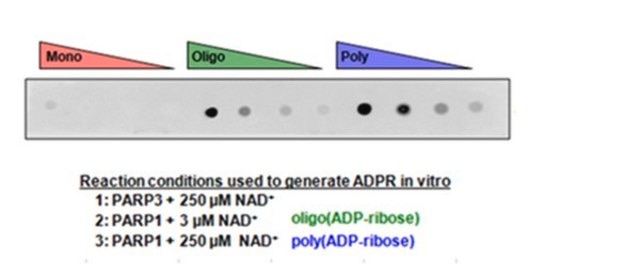
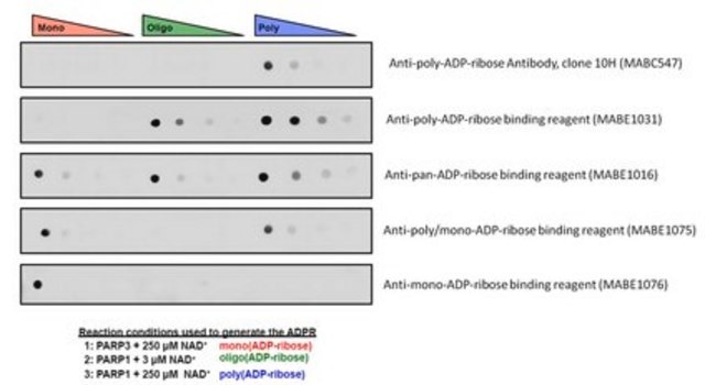
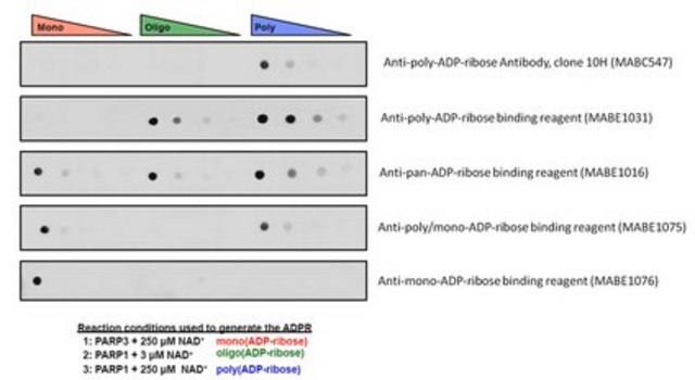
Dot Blot Specificity Analysis: This reagent detected mono(ADPR) on recombinant PARP3 protein, as well as mono(ADPR) and poly(ADPR) on recombinant PARP1 recombinant protein (Lee Kraus, University of Texas Southwestern Medical Center).
Immunoprecipitation Analysis: A representative lot of Anti-pan-ADP-ribose binding reagent immunoprecipitated ADP-ribosylated proteins from nuclear extract (Lee Kraus, University of Texas Southwestern).
Western Blotting Analysis: A representative lot detected auto-ADP-ribosylation activity of PARP1/2/3 and mutants in the presence of NAD+ or various NAD+ analogs (Gibson, B.A., et al. (2016). Science. 353(6294):45-50).
Western Blotting Analysis: A representative lot detected PARP1-catalyzed NELF-E ADP-ribosylation in cell-free enzymatic reactions as well as ADP-ribosylation of exogenously expressed FLAG-tagged NELF-E in HEK293T cells. PARP inhibitor PJ34 or P-TEFb/CDK9 inhibitor flavopiridol treatment decreased cellular NELF-E ADP-ribosylation level (Gibson, B.A., et al. (2016). Science. 353(6294):45-50).
Research Category
Epigenetics & Nuclear Function
Epigenetics & Nuclear Function
Research Sub Category
General Post-translation Modification
General Post-translation Modification
Quality
Evaluated by Western Blotting on ADP-ribosylated PARP1 and PARP3 recombinant proteins.
Western Blotting Analysis: This reagent detected mono(ADPR) on recombinant PARP3 protein, as well as mono(ADPR) and poly(ADPR) on recombinant PARP1 protein (Lee Kraus, University of Texas Southwestern Medical Center).
Western Blotting Analysis: This reagent detected mono(ADPR) on recombinant PARP3 protein, as well as mono(ADPR) and poly(ADPR) on recombinant PARP1 protein (Lee Kraus, University of Texas Southwestern Medical Center).
Target description
Variable depending on the target proteins and the extend of ADP-ribosylation.
Physical form
Format: Purified
Ni-NTA agarose
Purified from E. coli by Ni-NTA agarose. Supplied in buffer containing 10 mM Tris pH 7.5, 0.2 M NaCl, 10% Glycerol, 10 mM Imidazole, 1 mM PMSF, 1 mM β-Mercaptoethanol, 10% glycerol without preservatives.
Storage and Stability
Stable for 1 year at -80°C from date of receipt.
Handling Recommendations: Upon receipt and prior to removing the cap, centrifuge the vial and gently mix the solution. Aliquot into microcentrifuge tubes and store at -80°C. Avoid repeated freeze/thaw cycles, which may damage IgG and affect product performance.
Handling Recommendations: Upon receipt and prior to removing the cap, centrifuge the vial and gently mix the solution. Aliquot into microcentrifuge tubes and store at -80°C. Avoid repeated freeze/thaw cycles, which may damage IgG and affect product performance.
Other Notes
Concentration: Please refer to lot specific datasheet.
Disclaimer
Unless otherwise stated in our catalog or other company documentation accompanying the product(s), our products are intended for research use only and are not to be used for any other purpose, which includes but is not limited to, unauthorized commercial uses, in vitro diagnostic uses, ex vivo or in vivo therapeutic uses or any type of consumption or application to humans or animals.
Not finding the right product?
Try our Product Selector Tool.
wgk_germany
WGK 2
flash_point_f
Not applicable
flash_point_c
Not applicable
Certificates of Analysis (COA)
Search for Certificates of Analysis (COA) by entering the products Lot/Batch Number. Lot and Batch Numbers can be found on a product’s label following the words ‘Lot’ or ‘Batch’.
Already Own This Product?
Find documentation for the products that you have recently purchased in the Document Library.
Tao Qin et al.
Molecular plant, 12(9), 1243-1258 (2019-05-19)
Plasma membrane-associated abscisic acid (ABA) signal transduction is an integral part of ABA signaling. The C2-domain ABA-related (CAR) proteins play important roles in the recruitment of ABA receptors to the plasma membrane to facilitate ABA signaling. However, how CAR proteins
Karunakaran Kalesh et al.
Scientific reports, 9(1), 6655-6655 (2019-05-02)
ADP-ribosylation is integral to a diverse range of cellular processes such as DNA repair, chromatin regulation and RNA processing. However, proteome-wide investigation of its cellular functions has been limited due to numerous technical challenges including the complexity of the poly(ADP-ribose)
Daniel Roderer et al.
Nature communications, 10(1), 5263-5263 (2019-11-22)
Tc toxins are bacterial protein complexes that inject cytotoxic enzymes into target cells using a syringe-like mechanism. Tc toxins are composed of a membrane translocator and a cocoon that encapsulates a toxic enzyme. The toxic enzyme varies between Tc toxins
Katie Pollock et al.
Methods in molecular biology (Clifton, N.J.), 1608, 445-473 (2017-07-12)
The poly(ADP-ribose)polymerase (PARP) enzyme tankyrase (TNKS/ARTD5, TNKS2/ARTD6) uses its ankyrin repeat clusters (ARCs) to recognize degenerate peptide motifs in a wide range of proteins, thereby recruiting such proteins and their complexes for scaffolding and/or poly(ADP-ribosyl)ation. Here, we provide guidance for
Chao-Cheng Cho et al.
Nature communications, 10(1), 1491-1491 (2019-04-04)
Poly-ADP-ribosylation, a post-translational modification involved in various cellular processes, is well characterized in eukaryotes but thought to be devoid in bacteria. Here, we solve crystal structures of ADP-ribose-bound poly(ADP-ribose)glycohydrolase from the radioresistant bacterium Deinococcus radiodurans (DrPARG), revealing a solvent-accessible 2'-hydroxy
Our team of scientists has experience in all areas of research including Life Science, Material Science, Chemical Synthesis, Chromatography, Analytical and many others.
Contact Technical Service