MABF121
Anti-SV40 Large T Antigen Antibody, clone PAb416
clone PAb416, from mouse
Synonym(s):
Large T antigen, LT, LT-AG
Sign Into View Organizational & Contract Pricing
All Photos(1)
About This Item
UNSPSC Code:
12352203
eCl@ss:
32160702
NACRES:
NA.41
Recommended Products
biological source
mouse
Quality Level
antibody form
purified antibody
antibody product type
primary antibodies
clone
PAb416, monoclonal
species reactivity
SV40-infected cells
technique(s)
immunocytochemistry: suitable
western blot: suitable
isotype
IgG2aκ
NCBI accession no.
UniProt accession no.
shipped in
wet ice
target post-translational modification
unmodified
General description
Both the Simian virus (SV40) large T and small T antigens are encoded by the early region of the SV40 genome. The large T antigen binds DNA, and complexes with a 53,000 dalton cellular protein, p53, which is required for initiation of viral DNA replication during lytic growth. In addition the large T antigen binds DNA polymerase and the transcription factor AP-2 and forms a specific complex with the P105 product of the retinoblastoma susceptibility gene.
Specificity
This antibody recognizes the large T antigen of SV40.
Immunogen
Epitope: Large T antigen of SV40
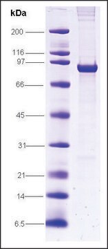
Purified large T-antigen of SV40
Application
Anti-SV40 Large T Antigen Antibody, clone PAb416 is an antibody against SV40 Large T Antigen for use in Western Blotting, ICC.
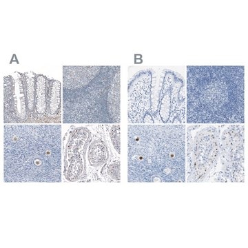
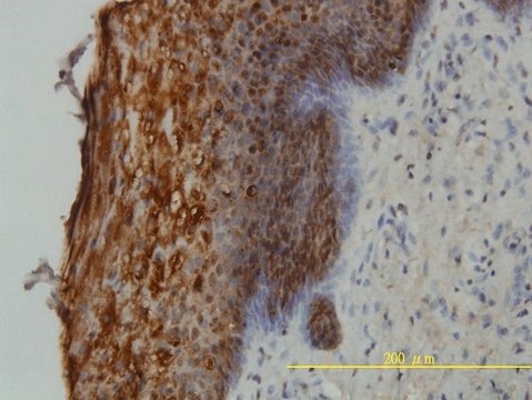
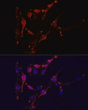
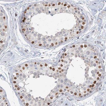
Immunocytochemistry Analysis: A representative lot from an independent laboratory detected SV40 Large T Antigen in ICC (Sabatier, J., et al. (2005). 58(4):429-431.; Del Valle, L., et al. (2004). J Virol. 78(7):3462-3469.).
Research Category
Inflammation & Immunology
Inflammation & Immunology
Research Sub Category
Immunoglobulins & Immunology
Immunoglobulins & Immunology
Quality
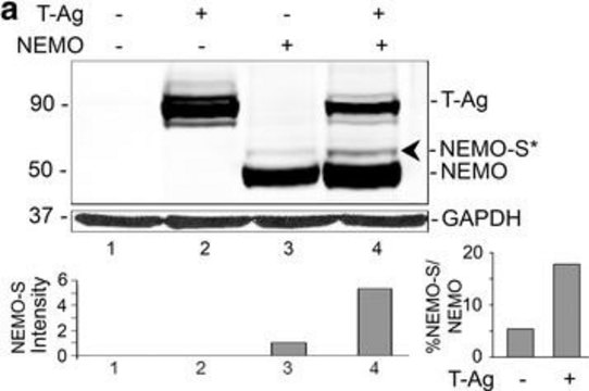
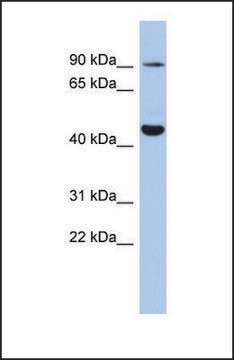
Evaluated by Western Blot in Cos-1 cell lysate.
Western Blot Analysis: 1 µg/mL of this antibody detected SV40 Large T Antigen in 10 µg of Cos-1 cell lysate.
Western Blot Analysis: 1 µg/mL of this antibody detected SV40 Large T Antigen in 10 µg of Cos-1 cell lysate.
Target description
~82 kDa observed
Physical form
Format: Purified
Protein G Purified
Purified mouse monoclonal IgG2aκ in buffer containing 0.1 M Tris-Glycine (pH 7.4), 150 mM NaCl with 0.05% sodium azide.
Storage and Stability
Stable for 1 year at 2-8°C from date of receipt.
Analysis Note
Control
Cos-1 cell lysate
Cos-1 cell lysate
Other Notes
Concentration: Please refer to the Certificate of Analysis for the lot-specific concentration.
Disclaimer
Unless otherwise stated in our catalog or other company documentation accompanying the product(s), our products are intended for research use only and are not to be used for any other purpose, which includes but is not limited to, unauthorized commercial uses, in vitro diagnostic uses, ex vivo or in vivo therapeutic uses or any type of consumption or application to humans or animals.
Not finding the right product?
Try our Product Selector Tool.
wgk_germany
WGK 1
flash_point_f
Not applicable
flash_point_c
Not applicable
Certificates of Analysis (COA)
Search for Certificates of Analysis (COA) by entering the products Lot/Batch Number. Lot and Batch Numbers can be found on a product’s label following the words ‘Lot’ or ‘Batch’.
Already Own This Product?
Find documentation for the products that you have recently purchased in the Document Library.
Immunodetection of SV40 large T antigen in human central nervous system tumours.
Sabatier, J, et al.
Journal of Clinical Pathology, 58, 429-431 (2005)
Primary central nervous system lymphoma expressing the human neurotropic polyomavirus, JC virus, genome.
Del Valle, Luis, et al.
Journal of virology, 78, 3462-3469 (2004)
Yasuko Orba et al.
The Journal of general virology, 92(Pt 4), 789-795 (2010-12-24)
To investigate polyomavirus infection in wild rodents, we analysed DNA samples from the spleens of 100 wild rodents from Zambia using a broad-spectrum PCR-based assay. A previously unknown polyomavirus genome was identified in a sample from a multimammate mouse (Mastomys
Ursula Neu et al.
PLoS pathogens, 9(10), e1003688-e1003688 (2013-10-17)
Viruses within a family often vary in their cellular tropism and pathogenicity. In many cases, these variations are due to viruses switching their specificity from one cell surface receptor to another. The structural requirements that underlie such receptor switching are
Furong Yuan et al.
Oncology reports, 23(2), 377-386 (2010-01-01)
Werner syndrome (WS) results from defects in the gene encoding WRN RecQ helicase. WS fibroblasts undergo premature senescence in culture. Because cellular senescence is a tumor suppressor mechanism, we examined whether WS fibroblasts exhibited reduced tumorigenicity, in comparison to control
Our team of scientists has experience in all areas of research including Life Science, Material Science, Chemical Synthesis, Chromatography, Analytical and many others.
Contact Technical Service