Skip to Content
MilliporeSigma
All Photos(1)

Documents

MABF33

Sigma-Aldrich

Anti-MHC class II (I-A/I-E) Antibody, clone M5/114

clone M5/114, from rat

Synonym(s):

CD74 molecule, major histocompatibility complex, class II invariant chain, Ia-associated invariant chain, CD74 antigen (invariant polypeptide of major histocompatibility complex, class II antigen-associated), gamma chain of class II antigens, Ia antigen-

Sign Into View Organizational & Contract Pricing


About This Item

UNSPSC Code:
12352203
eCl@ss:
32160702
NACRES:
NA.41

biological source

rat

Quality Level

antibody form

purified antibody

antibody product type

primary antibodies

clone

M5/114, monoclonal

species reactivity

mouse

technique(s)

flow cytometry: suitable
immunohistochemistry: suitable
western blot: suitable

isotype

IgG2bκ

NCBI accession no.

UniProt accession no.

shipped in

wet ice

target post-translational modification

unmodified

Gene Information

mouse ... Cd74(16149)

Related Categories

General description

MHC class II-associated invariant chain (I-A/I-E), also known as Ia antigen-associated invariant chain, are single-pass type II membrane glycoproteins containing a single thyroglobulin type-I domain. They are critical components in the processing of MHC class II antigen through forming a stabilizing complex following the alpha/beta heterodimer synthesis, and orchestrating complex trafficking away from the ER compartments in which class II peptide loading occurs.

Application

Detect MHC class II (I-A/I-E) using this Anti-MHC class II (I-A/I-E) Antibody, clone M5/114 validated for use in WB, FC, IH.
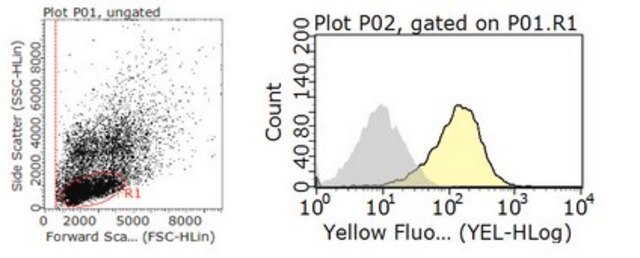
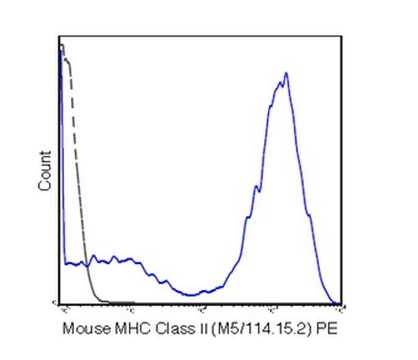
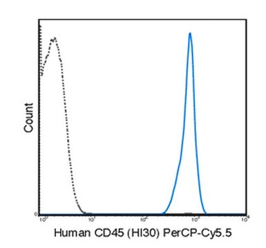
Flow Cytometry Analysis: A previous lot was used by an independent laboratory in FC on spleen cells. (Bhattacharya, A., et al. (1981). The Journal of immunology. 127(6):2488-2496.)

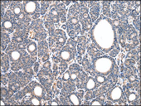
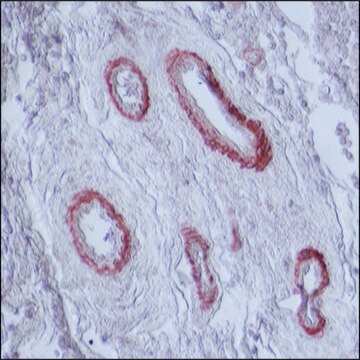
Immunohistochemistry Analysis: A previous lot was used by an independent laboratory in IH on thymus tissue. (de Bont, E., et al. (1999). Int. Immunol. 11(8):1295-1306.)

Quality

Evaluated by Western Blot in mouse spleen tissue lysate.

Western Blot Analysis: 1 µg/mL of this antibody detected MHC class II (I-A/I-E) in 10 µg of mouse spleen tissue lysate.

Target description

~32 kDa observed.
There are 2 isoforms produced by alternative splicing: 32 kDa Isoform Long and 24 kDa Isoform Short.

Physical form

Format: Purified

Other Notes

Concentration: Please refer to the Certificate of Analysis for the lot-specific concentration.

Not finding the right product?  

Try our Product Selector Tool.

wgk_germany

WGK 1

flash_point_f

Not applicable

flash_point_c

Not applicable


Certificates of Analysis (COA)

Search for Certificates of Analysis (COA) by entering the products Lot/Batch Number. Lot and Batch Numbers can be found on a product’s label following the words ‘Lot’ or ‘Batch’.

Already Own This Product?

Find documentation for the products that you have recently purchased in the Document Library.

Visit the Document Library

Libin Abraham et al.
Scientific reports, 7(1), 11379-11379 (2017-09-14)
Single-particle tracking (SPT) is a powerful method for exploring single-molecule dynamics in living cells with nanoscale spatiotemporal resolution. Photostability and bright fluorescence make quantum dots (Qdots) a popular choice for SPT. However, their large size could potentially alter the mobility
Circulating Porphyromonas gingivalis lipopolysaccharide resets cardiac homeostasis in mice through a matrix metalloproteinase-9-dependent mechanism.
Deleon-Pennell, KY; de Castro Bras, LE; Lindsey, ML
Physiological Reports null
Mahmoud Elashiry et al.
Journal of extracellular vesicles, 9(1), 1795362-1795362 (2020-09-19)
Chronic bone degenerative diseases represent a major threat to the health and well-being of the population, particularly those with advanced age. This study isolated exosomes (EXO), natural nano-particles, from dendritic cells, the "directors" of the immune response, to examine the
Madison Bolger-Munro et al.
eLife, 8 (2019-06-04)
When B cells encounter antigens on the surface of an antigen-presenting cell (APC), B cell receptors (BCRs) are gathered into microclusters that recruit signaling enzymes. These microclusters then move centripetally and coalesce into the central supramolecular activation cluster of an
Jia C Wang et al.
Journal of cell science, 130(6), 1094-1109 (2017-02-09)
B cells that bind antigens displayed on antigen-presenting cells (APCs) form an immune synapse, a polarized cellular structure that optimizes the dual functions of the B cell receptor (BCR), signal transduction and antigen internalization. Immune synapse formation involves polarization of

Our team of scientists has experience in all areas of research including Life Science, Material Science, Chemical Synthesis, Chromatography, Analytical and many others.

Contact Technical Service