Skip to Content
MilliporeSigma
All Photos(1)

Documents

MABF512

Sigma-Aldrich

Anti-CD11b Antibody (human/mouse), APC-Cy7, clone M1/70

clone M1/70, 0.2 mg/mL, from rat

Synonym(s):

Integrin alpha-M, CD11 antigen-like family member B, CR-3 alpha chain, Cell surface glycoprotein MAC-1 subunit alpha, Leukocyte adhesion receptor MO1, Neutrophil adherence receptor, CD antigen CD11b

Sign Into View Organizational & Contract Pricing


About This Item

UNSPSC Code:
12352203
eCl@ss:
32160702
NACRES:
NA.41

biological source

rat

Quality Level

conjugate

APC-Cy7

antibody form

purified antibody

antibody product type

primary antibodies

clone

M1/70, monoclonal

species reactivity

mouse, human

packaging

antibody small pack of 25 μg

concentration

0.2 mg/mL

technique(s)

flow cytometry: suitable
immunofluorescence: suitable
immunohistochemistry: suitable
immunoprecipitation (IP): suitable

isotype

IgG2bκ

UniProt accession no.

shipped in

wet ice

target post-translational modification

unmodified

Gene Information

human ... ITGAM(3684)

Related Categories

General description

The M1/70 antibody reacts with human and mouse CD11b, also known as integrin alpha M. This 165-170 kDa cell surface glycoprotein is part of a family of integrin receptors that mediate adhesion between cells (cell-cell) and components of the extracellular matrix, e.g. fibrinogen (cell-matrix). In addition, integrins are active signaling receptors which recruit leukocytes to inflammatory sites and promote cell activation. Complete, functional integrin receptors consist of distinct combinations of integrin chains which are differentially expressed. Integrin alpha M (CD11b) assembles with Integrin beta-2 (CD18) into a receptor known as Macrophage Antigen-1 (Mac-1) or complement receptor type 3 (CR3). This receptor binds and induces intracellular signaling through ICAM-1 on endothelial cells and can also facilitate removal of iC3b bearing foreign cells.The M1/70 antibody is widely used as a marker for CD11b expression on mouse macrophages, granulocytes, neutrophils, and NK cells. The antibody is also reported to be cross-reactive for Rhesus macaque CD11b.

Immunogen

Immunogen information for this antibody is proprietary information.

Application

Research Category
Inflammation & Immunology
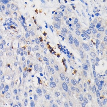
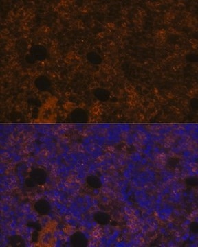
This rat monoclonal antibody Anti-CD11b (human/mouse), APC-Cy7, clone M1/70 validated for use in IP, IHC, Flow Cytometry & Immunofluorescence for the detection of CD11b.

Quality

The monoclonal antibody was purified from tissue culture supernatant via affinity chromatography. The purified antibody was conjugated under optimal conditions, with unreacted dye removed from the preparation.

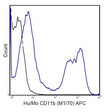
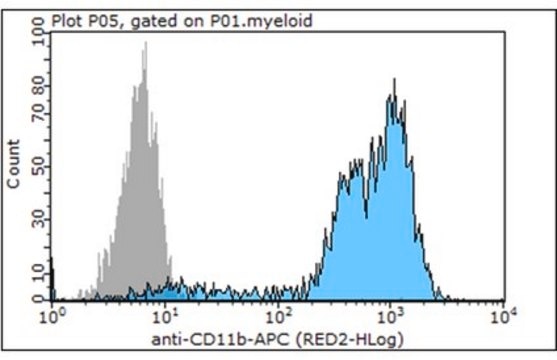
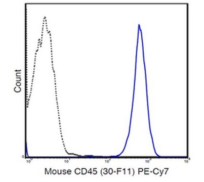
Flow Cytometry Analysis: 0.125 μg of this antibody detected CD11b in one million C57Bl/6 bone marrow cells.

Physical form

Purified Rat Monoclonal Antibody in buffer containing 10 mM NaH2PO4, 150 mM NaCl, 0.1% gelatin, pH 7.2, and up to 0.1% sodium azide.
Size Exclusion Chromatography

Storage and Stability

It is recommended to store the product undiluted at 2-8°C and protected from prolonged exposure to light. Do not freeze.

Disclaimer

Unless otherwise stated in our catalog or other company documentation accompanying the product(s), our products are intended for research use only and are not to be used for any other purpose, which includes but is not limited to, unauthorized commercial uses, in vitro diagnostic uses, ex vivo or in vivo therapeutic uses or any type of consumption or application to humans or animals.

Not finding the right product?  

Try our Product Selector Tool.

wgk_germany

nwg

flash_point_f

Not applicable

flash_point_c

Not applicable


Certificates of Analysis (COA)

Search for Certificates of Analysis (COA) by entering the products Lot/Batch Number. Lot and Batch Numbers can be found on a product’s label following the words ‘Lot’ or ‘Batch’.

Already Own This Product?

Find documentation for the products that you have recently purchased in the Document Library.

Visit the Document Library

Ileana Ruxandra Botusan et al.
PloS one, 13(3), e0193084-e0193084 (2018-03-14)
IGF-I is a growth factor, which is expressed in virtually all tissues. The circulating IGF-I is however derived mainly from the liver. IGF-I promotes wound healing and its levels are decreased in wounds with low regenerative potential such as diabetic

Our team of scientists has experience in all areas of research including Life Science, Material Science, Chemical Synthesis, Chromatography, Analytical and many others.

Contact Technical Service