MABF977
Anti-ICOS/CD278 Antibody, clone C398.4A
clone C398.4A, from hamster(Armenian)
Synonym(s):
Inducible T-cell costimulator, Activation-inducible lymphocyte immunomediatory molecule, CCLP, CD278, CD28 and CTLA-4-like protein, CD28-related protein 1, CRP-1, H4
About This Item
Recommended Products
biological source
hamster (Armenian)
Quality Level
antibody form
purified immunoglobulin
antibody product type
primary antibodies
clone
C398.4A, monoclonal
species reactivity
mouse, human
species reactivity (predicted by homology)
rhesus macaque (based on 100% sequence homology)
technique(s)
flow cytometry: suitable
immunocytochemistry: suitable
immunohistochemistry: suitable
immunoprecipitation (IP): suitable
isotype
IgG
NCBI accession no.
UniProt accession no.
shipped in
ambient
target post-translational modification
unmodified
Gene Information
human ... ICOS(29851)
mouse ... Icos(54167)
Related Categories
General description
Specificity
Immunogen
Application
Flow Cytometry Analysis: A representative lot detected the surface expression of exogenously transfected human ICOS on the surface of murine L cell fibroblasts. Clone C398.4A and another ICOS mAb (clone F44) competed against each other for the the suface staining of PHA-activated human peripheral blood T-cells (Buonfiglio, D., et al. (2000). Eur. J. Immunol. 30(12):3463-3467).
Flow Cytometry Analysis: A representative lot detected a greatly enhanced ICOS/CD278- (hpH4; human putative mouse H4 homolog) positive cell population among isolated human PBMCs following PHA stimulation (Buonfiglio, D., et al. (1999). Eur. J. Immunol. 29(9):2863-2874).
Flow Cytometry Analysis: A representative lot immunostained D10.G4.1 murine TH2 cells as well as activated, but not resting, T and B cells freshly isolated from mouse spleen (Redoglia, V., et al. (1996). Eur. J. Immunol. 26(11):2781-2789).
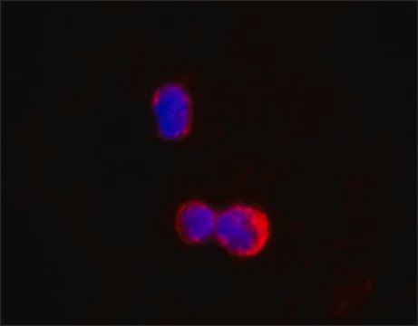
Immunocytochemistry Analysis: A representative lot immunostained H4 (ICOS/CD278) immunoreactivity co-capped with that of TCR mAb 3D3 on the surface of D10.G4.1 murine TH2 cells by immunofluorescence microscopy (Redoglia, V., et al. (1996). Eur. J. Immunol. 26(11):2781-2789).
Immunohistochemistry Analysis: A representative lot immunostained the membrane of hpH4- (ICOS/CD278) positive lymphocytes in acetone-fixed frozen human reactive lymph node sections, while most neoplastic cells showed cytoplasmic hpH4 staining in frozen PTCL-U (peripheral T cell lymphoma, unspecified) and angioimmunoblastic T cell lymphoma sections (Buonfiglio, D., et al. (1999). Eur. J. Immunol. 29(9):2863-2874).
Immunoprecipitation Analysis: A representative lot immunoprecipitated and depleted ICOS from human T-cell lymphoma HuT 78 cell lysates (Buonfiglio, D., et al. (2000). Eur. J. Immunol. 30(12):3463-3467).
Immunoprecipitation Analysis: A representative lot immunoprecipitated H4 (ICOS/CD278) from PHA-activiated human peripheral blood (PB) T cells in a ~55-70 kDa glycosylated dimeric form (Buonfiglio, D., et al. (1999). Eur. J. Immunol. 29(9):2863-2874).
Immunoprecipitation Analysis: A representative lot immunoprecipitated H4 (ICOS/CD278) from D10.G4.1 murine TH2 cells in a glycosylated dimeric form (Redoglia, V., et al. (1996). Eur. J. Immunol. 26(11):2781-2789).
Inflammation & Immunology
Quality
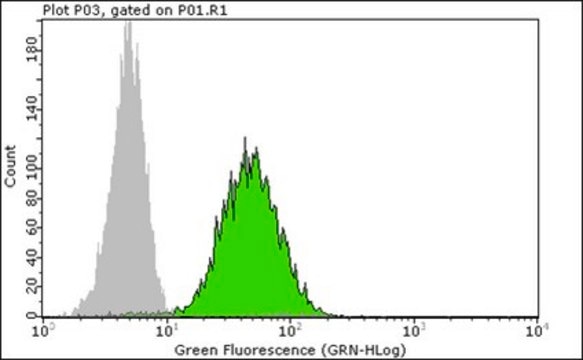
Flow Cytometry Analysis: 0.1 µg of this antibody detected ICOS/CD278 immunoreactivity on the surface of PHA-activated (5 µg/mL at 37°C for 72 hours) human PBMCs.
Target description
Physical form
Storage and Stability
Handling Recommendations: Upon receipt and prior to removing the cap, centrifuge the vial and gently mix the solution. Aliquot into microcentrifuge tubes and store at -20°C. Avoid repeated freeze/thaw cycles, which may damage IgG and affect product performance.
Other Notes
Disclaimer
Not finding the right product?
Try our Product Selector Tool.
wgk_germany
WGK 2
flash_point_f
Not applicable
flash_point_c
Not applicable
Certificates of Analysis (COA)
Search for Certificates of Analysis (COA) by entering the products Lot/Batch Number. Lot and Batch Numbers can be found on a product’s label following the words ‘Lot’ or ‘Batch’.
Already Own This Product?
Find documentation for the products that you have recently purchased in the Document Library.
Our team of scientists has experience in all areas of research including Life Science, Material Science, Chemical Synthesis, Chromatography, Analytical and many others.
Contact Technical Service