MABN466
Anti-Ankyrin-G Antibody, clone N106/36
clone N106/36, from mouse
Synonym(s):
Ankyrin-3, ANK-3, Ankyrin-G
Sign Into View Organizational & Contract Pricing
All Photos(2)
About This Item
UNSPSC Code:
12352203
eCl@ss:
32160702
NACRES:
NA.41
Recommended Products
biological source
mouse
Quality Level
antibody form
purified immunoglobulin
antibody product type
primary antibodies
clone
N106/36, monoclonal
species reactivity
rat
technique(s)
immunohistochemistry: suitable
isotype
IgG2aκ
NCBI accession no.
UniProt accession no.
shipped in
wet ice
target post-translational modification
unmodified
Gene Information
rat ... Ank3(361833)
Related Categories
General description
Ankyrins are a family of spectrin-binding proteins that couple the spectrin/actin cytoskeletal network to the cytoplasmic domains of integral membrane proteins which include channels such as the anion exchanger, voltage-dependent sodium channel, and the Na/K ATPase, as well as neuronal cell adhesion molecules related to L1. In the nervous system, three different ankyrins are known to exist; ankyrinB, ankyrinG, and ankyrinR. All three ankyrins contain an N-terminal membrane binding domain, an internal domain capable of binding spectrin, and a C-terminal regulatory domain which is the target of alternative splicing. AnkyrinB, the predominant neuronal ankyrin includes two isoforms of ~220 kDa and ~440 kDa. The ~220 kDa alternatively spliced isoform is the major form present in the adult brain, while the ~440 kDa form is preferentially expressed in the developing neonatal brain. Three forms of ankyrinG (~190 kDa, ~270 kDa, ~480 kDa) are localized in axonal initial segments and the nodes of Ranvier of myelinated axons where fluxes of action potential occur and ion channels are enriched. Ankyrins comprise ~0.5%-1% of the total membrane protein in the adult vertebrate brain.
Specificity
Other homologies: Human (92% sequence homology), and Mouse (87% sequence homology).
Immunogen
Recombinant protein corresponding to human Ankyrin-G.
Application
Anti-Ankyrin-G Antibody, clone N106/36 is a highly specific mouse monoclonal antibody & that targets Ankyrin & has been tested in IHC.
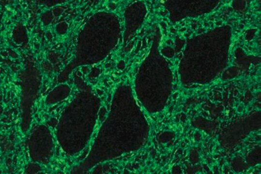
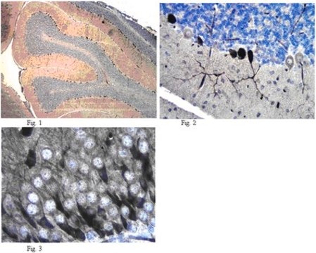
Immunohistochemistry Analysis: A 1:2,000 dilution from a representative lot detected Ankyrin-G in rat cerebral cortex tissue.
Immunohistochemistry Analysis: A representative lot detected Ankyrin-G in rat optic nerve tissue.
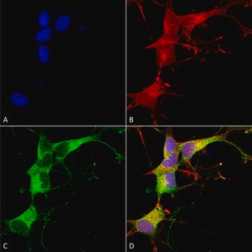
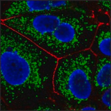
Immunofluorescence Analysis: A representative lot detected Ankyrin-G in adult rat cortex tissue.
Immunofluorescence Analysis: A representative lot from an independent laboratory detected Ankyrin-G in a rat model of TLE mEC layer II neurons (Hargus, N. J., et al. (2011). Neurobiol Dis. 41(2):361-376.)
Research Category
Neuroscience
Neuroscience
Research Sub Category
Developmental Neuroscience
Developmental Neuroscience
Quality
Evaluated by Immunohistochemistry in rat hippocampus tissue.
Immunohistochemistry Analysis: A 1:500 dilution from a representative lot detected Ankyrin-G in rat hippocampus tissue.
Immunohistochemistry Analysis: A 1:500 dilution from a representative lot detected Ankyrin-G in rat hippocampus tissue.
Target description
480 kDa calculated
Physical form
Format: Purified
Protein G Purified
Purified mouse monoclonal IgG2a κ in 0.1 M Tris-Glycine (pH 7.4), 150 mM NaCl with 0.05% sodium azide.
Storage and Stability
Stable for 1 year at 2-8°C from date of receipt.
Analysis Note
Control
Rat hippocampus tissue
Rat hippocampus tissue
Other Notes
Concentration: Please refer to the Certificate of Analysis for the lot-specific concentration.
Disclaimer
Unless otherwise stated in our catalog or other company documentation accompanying the product(s), our products are intended for research use only and are not to be used for any other purpose, which includes but is not limited to, unauthorized commercial uses, in vitro diagnostic uses, ex vivo or in vivo therapeutic uses or any type of consumption or application to humans or animals.
Not finding the right product?
Try our Product Selector Tool.
wgk_germany
WGK 1
flash_point_f
Not applicable
flash_point_c
Not applicable
Certificates of Analysis (COA)
Search for Certificates of Analysis (COA) by entering the products Lot/Batch Number. Lot and Batch Numbers can be found on a product’s label following the words ‘Lot’ or ‘Batch’.
Already Own This Product?
Find documentation for the products that you have recently purchased in the Document Library.
Tuancheng Feng et al.
Acta neuropathologica communications, 10(1), 33-33 (2022-03-16)
TMEM106B, a type II lysosomal transmembrane protein, has recently been associated with brain aging, hypomyelinating leukodystrophy, frontotemporal lobar degeneration (FTLD) and several other brain disorders. TMEM106B is critical for proper lysosomal function and TMEM106B deficiency leads to myelination defects, FTLD
Amanda M Brown et al.
Scientific reports, 9(1), 1742-1742 (2019-02-12)
Purkinje cells receive synaptic input from several classes of interneurons. Here, we address the roles of inhibitory molecular layer interneurons in establishing Purkinje cell function in vivo. Using conditional genetics approaches in mice, we compare how the lack of stellate
Kelsie Eichel et al.
Nature, 609(7925), 128-135 (2022-08-18)
Neurons are highly polarized cells that face the fundamental challenge of compartmentalizing a vast and diverse repertoire of proteins in order to function properly1. The axon initial segment (AIS) is a specialized domain that separates a neuron's morphologically, biochemically and
Mable Lam et al.
Nature communications, 13(1), 5583-5583 (2022-09-24)
Myelin is required for rapid nerve signaling and is emerging as a key driver of CNS plasticity and disease. How myelin is built and remodeled remains a fundamental question of neurobiology. Central to myelination is the ability of oligodendrocytes to
Microglial Displacement of GABAergic Synapses Is a Protective Event during Complex Febrile Seizures.
Yushan Wan et al.
Cell reports, 33(5), 108346-108346 (2020-11-05)
Complex febrile seizures (FSs) lead to a high risk of intractable temporal lobe epilepsy during adulthood, yet the pathological process of complex FSs is largely unknown. Here, we demonstrate that activated microglia extensively associated with glutamatergic neuronal soma displace surrounding
Our team of scientists has experience in all areas of research including Life Science, Material Science, Chemical Synthesis, Chromatography, Analytical and many others.
Contact Technical Service