Skip to Content
MilliporeSigma
All Photos(2)

Documents

MABN473

Sigma-Aldrich

Anti-TMEM106B Antibody, clone TME-N 6F2

clone TME-N 6F2, from rat

Synonym(s):

Transmembrane protein 106B, TMEM106B

Sign Into View Organizational & Contract Pricing


About This Item

UNSPSC Code:
12352203
eCl@ss:
32160702
NACRES:
NA.41

biological source

rat

Quality Level

antibody form

purified immunoglobulin

antibody product type

primary antibodies

clone

TME-N 6F2, monoclonal

species reactivity

rat, human

technique(s)

immunofluorescence: suitable
western blot: suitable

NCBI accession no.

UniProt accession no.

shipped in

wet ice

target post-translational modification

unmodified

Gene Information

General description

Transmembrane protein 106B (TMEM106B) is a single-pass type II membrane protein present in endosomal and lysosomal membranes, and is highly expressed in the frontal cortex. Increased TMEM106B expression in the brain may be linked to mechanisms of disease in frontotemporal lobar degeneration with TAR DNA-binding protein (TDP-43) inclusions (FTLD-TDP). Frontotemporal lobar degeneration (FTLD) is the second most common cause of presenile dementia.

Specificity

This antibody recognizes the N-terminus of TMEM106B.

Immunogen

Epitope: N-terminus
Recombinant protein corresponding to the N-terminus of human TMEM106B.

Application

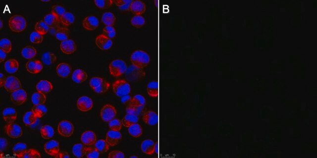
Anti-TMEM106B, clone TME-N 6F2 detects levels of TMEM106B proteins & has been published & validated for use in WB & IF.
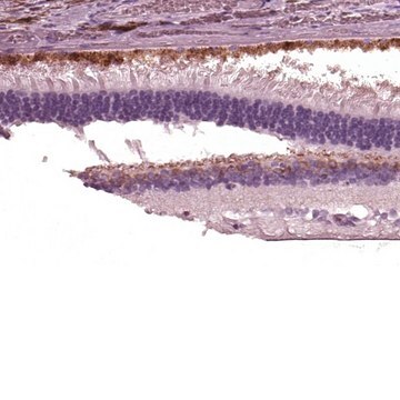
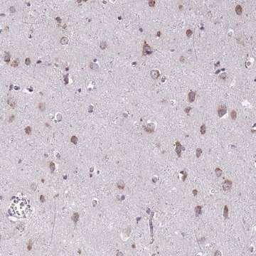
Immunofluorescence Analysis: A 1:50 dilution from a representative lot detected TMEM106B in rat cerebellum tissue.
Research Category
Neuroscience
Research Sub Category
Neurodegenerative Diseases

Quality

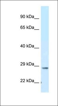
Evaluated by Western Blot in human cortex tissue lysate.

Western Blot Analysis: 1 µg/mL of this antibody detected TMEM106B in 10 µg of human cortex tissue lysate.

Target description

~30 kDa observed (At higher antibody concentrations, this protein may be observed at ~45 kDa). This protein may be observed at ~50 kDa due to glycosylation (Lang, C. M., et al. (2012). J Biol Chem. 287(23):19355-19365.).

Physical form

Format: Purified
Protein G Purified
Purified rat monoclonal IgG2c in buffer containing 0.1 M Tris-Glycine (pH 7.4), 150 mM NaCl with 0.05% sodium azide.

Storage and Stability

Stable for 1 year at 2-8°C from date of receipt.

Analysis Note

Control
Human cortex tissue lysate

Other Notes

Concentration: Please refer to the Certificate of Analysis for the lot-specific concentration.

Disclaimer

Unless otherwise stated in our catalog or other company documentation accompanying the product(s), our products are intended for research use only and are not to be used for any other purpose, which includes but is not limited to, unauthorized commercial uses, in vitro diagnostic uses, ex vivo or in vivo therapeutic uses or any type of consumption or application to humans or animals.

Not finding the right product?  

Try our Product Selector Tool.

wgk_germany

WGK 1

flash_point_f

Not applicable

flash_point_c

Not applicable


Certificates of Analysis (COA)

Search for Certificates of Analysis (COA) by entering the products Lot/Batch Number. Lot and Batch Numbers can be found on a product’s label following the words ‘Lot’ or ‘Batch’.

Already Own This Product?

Find documentation for the products that you have recently purchased in the Document Library.

Visit the Document Library

Christina M Lang et al.
The Journal of biological chemistry, 287(23), 19355-19365 (2012-04-19)
TMEM106B was identified as a major risk factor in a genome-wide association study for frontotemporal lobar degeneration (FTLD) with TAR DNA-binding protein (TDP)-43 pathology. The most significant association of TMEM106B single nucleotide polymorphisms with risk of FTLD-TDP was observed in
Benjamin M Schwenk et al.
The EMBO journal, 33(5), 450-467 (2013-12-21)
TMEM106B is a major risk factor for frontotemporal lobar degeneration with TDP-43 pathology. TMEM106B localizes to lysosomes, but its function remains unclear. We show that TMEM106B knockdown in primary neurons affects lysosomal trafficking and blunts dendritic arborization. We identify microtubule-associated
Common pathobiochemical hallmarks of progranulin-associated frontotemporal lobar degeneration and neuronal ceroid lipofuscinosis.
Gotzl, JK; Mori, K; Damme, M; Fellerer, K; Tahirovic, S; Kleinberger, G; Janssens et al.
Acta neuropathologica null

Our team of scientists has experience in all areas of research including Life Science, Material Science, Chemical Synthesis, Chromatography, Analytical and many others.

Contact Technical Service