MABS1916
Anti-c-Met Antibody, clone seeMet 13
clone seeMet 13, from mouse
Synonym(s):
Hepatocyte growth factor receptor, HGF receptor, HGF/SF receptor, Proto-oncogene c-Met, Scatter factor receptor, SF receptor, Tyrosine-protein kinase Met
About This Item
Recommended Products
biological source
mouse
Quality Level
antibody form
purified immunoglobulin
antibody product type
primary antibodies
clone
seeMet 13, monoclonal
species reactivity
human
technique(s)
flow cytometry: suitable
immunocytochemistry: suitable
immunoprecipitation (IP): suitable
inhibition assay: suitable
isotype
IgG1κ
NCBI accession no.
UniProt accession no.
shipped in
ambient
target post-translational modification
unmodified
Gene Information
human ... HGF(3082)
Related Categories
General description
Specificity
Immunogen
Application
Flow Cytometry Analysis: A representative lot immunostained live SNU-5 cells. A slight decrease in antibody immunoreactivity was observed when the temperature was dropped from 37°C to 4°C (Wong, J.S., et al. (2013). Oncotarget. 4(7):1019-1036).
Immunocytochemistry Analysis: A representative lot immunostained live SNU-5 cells. Indirect fluorescence labelling following subsequent cell fixation and permeabilization revealed increased antibody cytoplasmic internalization when antibody incubation was performed at 37°C than at 4°C (Wong, J.S., et al. (2013). Oncotarget. 4(7):1019-1036).
Immunoprecipitation Analysis: A representative lot immunoprecipitated c-Met from SNU-5 cell lysate (Wong, J.S., et al. (2013). Oncotarget. 4(7):1019-1036).
Inhibition Analysis: A representative lot prevented HGF-induced scatter of HaCaT cells. Clone seeMet 13 inhibited cell proliferation by blocking cell division without inducing apoptosis (Wong, J.S., et al. (2013). Oncotarget. 4(7):1019-1036).
Note: seeMet 13 exhibited low reactivity and specificity towards denatured c-Met by Western blotting. This monoclonal antibody is not recommended for Western blotting application (Wong, J.S., et al. (2013). Oncotarget. 4(7):1019-1036).
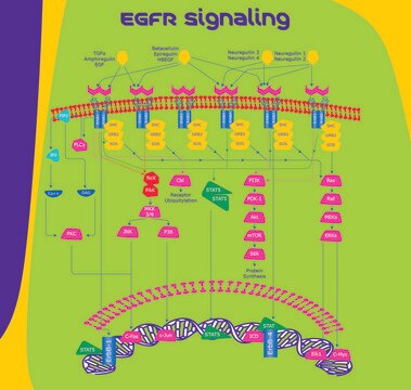
Signaling
Quality
Immunocytochemistry Analysis: A 1:500 dilution of this antibody detected both surface and internalized c-Met by fluorescent immunocytochemistry staining of 4% paraformaldehyde-fixed, 0.3% Triton X-100-permeabilized SNU-5 cells.
Target description
Physical form
Storage and Stability
Handling Recommendations: Upon receipt and prior to removing the cap, centrifuge the vial and gently mix the solution. Aliquot into microcentrifuge tubes and store at -20°C. Avoid repeated freeze/thaw cycles, which may damage IgG and affect product performance.
Other Notes
Disclaimer
Not finding the right product?
Try our Product Selector Tool.
wgk_germany
WGK 2
Certificates of Analysis (COA)
Search for Certificates of Analysis (COA) by entering the products Lot/Batch Number. Lot and Batch Numbers can be found on a product’s label following the words ‘Lot’ or ‘Batch’.
Already Own This Product?
Find documentation for the products that you have recently purchased in the Document Library.
Our team of scientists has experience in all areas of research including Life Science, Material Science, Chemical Synthesis, Chromatography, Analytical and many others.
Contact Technical Service
