General description
Tetraspanin-8 (UniProt: Q8R3G9; also known as Tspan8, Tspan-8) is encoded by the Tspan8 gene (Gene ID: 216350) in murine species. Tspan-8 is a multi-pass membrane protein of the tetraspanin family that is involved in the regulation of cell activation, growth, and motility. Members of this family have emerged as important regulators of expression levels, trafficking or post-translational modification of laminin-binding integrins. Tetraspanins are shown to associate with one another and cluster dynamically with a large variety of transmembrane and signal-transducing partners, forming specialized membrane microdomains called tetraspanin web. Tspan-8 is reported to be overexpressed during the progression of colorectal, liver, pancreatic and gastric cancers and its increased expression is shown to promote liver and lung metastasis. Overexpression of Tspan-8 has also been correlated with poor prognosis in human colorectal cancer. In metastatic pancreatic and colorectal carcinoma cell lines, PKC activation enhances the colocalization of Tspan-8 and Tspan CD151 with integrin 6 4 and subsequent internalization of this integrin-tetraspanin complex, resulting in decreased cell-matrix adhesion and increased cell migration. Tspan-8 knockdown in HT29 colon cancer cells is reported to significantly reduce their migratory ability as a result of an upregulation of both integrin-dependent cell-matrix adhesion and calcium-dependent cell-cell adhesion. (Ref.: Kharbili, ME., et al. (2017). Oncotarget. 8(10); 17140-17155).
Specificity
Clone 51/15-8G4-17-42-1 is a rat monoclonal antibody that detects human and murine Tetraspanin-8. It targets an epitope with in 12 amino acids from the extracellular domain.
Immunogen
Epitope: extracellular domain
KLH-conjugated linear peptide corresponding to 12 amino acids from the extracellular domain of mouse Tetraspanin-8.
Application
Anti-Tspan8-C, clone 51/15-8G4-17-42-1, Cat. No. MABT1351, is a rat monoclonal antibody that detects Tetraspanin-8 and has been tested for use in Immunohistochemistry and Western Blotting.
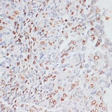
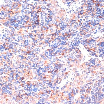
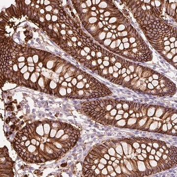
Immunohistochemistry Analysis: A representative lot detected Tspan8-C in Immunohistochemistry applications (Fu, N.Y., et. al. (2017). Nat Cell Biol. 19(3):164-176).
Research Category
Cell Structure
Quality
Evaluated by Western Blotting in HT-29 cell lysate.
Western Blotting Analysis: A 1:250 dilution of this antibody detected Tspan8-C in HT-29 cell lysate.
Target description
~25 kDa observed; 25.58 kDa calculated. Uncharacterized bands may be observed in some lysate(s).
Physical form
Format: Purified
Protein G purified
Purified rat monoclonal antibody IgG2a in buffer containing 0.1 M Tris-Glycine (pH 7.4), 150 mM NaCl with 0.05% sodium azide.
Storage and Stability
Stable for 1 year at 2-8°C from date of receipt.
Other Notes
Concentration: Please refer to lot specific datasheet.
Disclaimer
Unless otherwise stated in our catalog or other company documentation accompanying the product(s), our products are intended for research use only and are not to be used for any other purpose, which includes but is not limited to, unauthorized commercial uses, in vitro diagnostic uses, ex vivo or in vivo therapeutic uses or any type of consumption or application to humans or animals.
