Skip to Content
MilliporeSigma
All Photos(2)

Documents

MABT95

Sigma-Aldrich

Anti-Arp2/3 complex Antibody, clone 13C9

clone 13C9, from mouse

Synonym(s):

Actin-related protein 2/3 complex subunit 3, Arp2/3 complex 21 kDa subunit, p21-ARC

Sign Into View Organizational & Contract Pricing


About This Item

UNSPSC Code:
12352203
eCl@ss:
32160702
NACRES:
NA.41

biological source

mouse

Quality Level

antibody form

purified immunoglobulin

antibody product type

primary antibodies

clone

13C9, monoclonal

species reactivity

human

technique(s)

immunohistochemistry: suitable (paraffin)
western blot: suitable

isotype

IgG2aκ

NCBI accession no.

UniProt accession no.

shipped in

wet ice

target post-translational modification

unmodified

Gene Information

human ... ARPC3(10094)

General description

The Arp2/3 complex, which contains ARP2, ARP3, ARPC1B/p41-ARC, ARPC2/p34-ARC, ARPC3/p21-ARC, ARPC4/p20-ARC and ARPC5/p16-ARC, plays a major role in the regulation of the actin polymerization in the cytoskeleton. Two of complex′s seven subunits (ARP2 and ARP3) closely resemble monomeric actin and serve as nucleation sites for new actin filaments. The Arp2/3 complex works in conjunction with an activating nucleation-promoting factor (NPF) to aid in the formation of branched actin networks. The regulation of actin cytoskeleton reorganization is important for processes like cell migration, phagocytosis, and intracellular motility of lipid vesicles.

Immunogen

GST-tagged recombinant protein corresponding to human Arp2/3 complex.

Application

Anti-Arp2/3 complex Antibody, clone 13C9 detects level of Arp2/3 complex & has been published & validated for use in WB, IH(P).
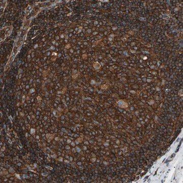
Immunohistochemistry Analysis: A 1:5,000 dilution from a representative lot detected Arp2/3 complex in human colorectal cancer and human colon tissues (antibody may need further dilution if staining is too strong - optimal dilution to be determined by the end user).
Research Category
Cell Structure
Research Sub Category
Cytoskeleton

Quality

Evaluated by Western Blot in MCF-7 cell lysate.

Western Blot Analysis: A 1:5,000 dilution of this antibody detected Arp2/3 complex on 10 µg of MCF-7 cell lysate.

Target description

~20 kDa observed

Physical form

Format: Purified
Protein G Purified
Purified mouse monoclonal IgG2aκ in buffer containing 0.1 M Tris-Glycine (pH 7.4), 150 mM NaCl with 0.05% sodium azide.

Storage and Stability

Stable for 1 year at 2-8°C from date of receipt.

Analysis Note

Control
MCF-7 cell lysate

Disclaimer

Unless otherwise stated in our catalog or other company documentation accompanying the product(s), our products are intended for research use only and are not to be used for any other purpose, which includes but is not limited to, unauthorized commercial uses, in vitro diagnostic uses, ex vivo or in vivo therapeutic uses or any type of consumption or application to humans or animals.

Not finding the right product?  

Try our Product Selector Tool.

wgk_germany

WGK 1

flash_point_f

Not applicable

flash_point_c

Not applicable


Certificates of Analysis (COA)

Search for Certificates of Analysis (COA) by entering the products Lot/Batch Number. Lot and Batch Numbers can be found on a product’s label following the words ‘Lot’ or ‘Batch’.

Already Own This Product?

Find documentation for the products that you have recently purchased in the Document Library.

Visit the Document Library

Aida M Lopez-Guerrero et al.
Scientific reports, 10(1), 6580-6580 (2020-04-22)
Tumor invasion requires efficient cell migration, which is achieved by the generation of persistent and polarized lamellipodia. The generation of lamellipodia is supported by actin dynamics at the leading edge where a complex of proteins known as the WAVE regulatory
Diana C Muñoz-Lasso et al.
Scientific reports, 10(1), 5207-5207 (2020-04-07)
Abnormalities in actin cytoskeleton have been linked to Friedreich's ataxia (FRDA), an inherited peripheral neuropathy characterised by an early loss of neurons in dorsal root ganglia (DRG) among other clinical symptoms. Despite all efforts to date, we still do not
Sarah R Barger et al.
Nature communications, 10(1), 1249-1249 (2019-03-21)
Phagocytosis of invading pathogens or cellular debris requires a dramatic change in cell shape driven by actin polymerization. For antibody-covered targets, phagocytosis is thought to proceed through the sequential engagement of Fc-receptors on the phagocyte with antibodies on the target

Our team of scientists has experience in all areas of research including Life Science, Material Science, Chemical Synthesis, Chromatography, Analytical and many others.

Contact Technical Service