MCD8MAG-48K
MILLIPLEX® Mouse CD8+ T Cell Magnetic Bead Panel - Immunology Multiplex Assay
Inflammation/Immunology Bead-Based Multiplex Assays using the Luminex technology enables the simultaneous analysis of multiple cytokine and chemokine CD8+ biomarkers in mouse serum, plasma and cell culture samples.
About This Item
Recommended Products
Quality Level
species reactivity
mouse
manufacturer/tradename
Milliplex®
assay range
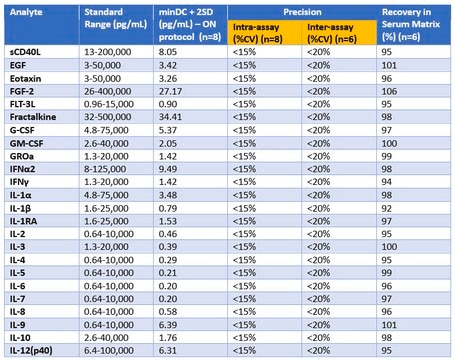
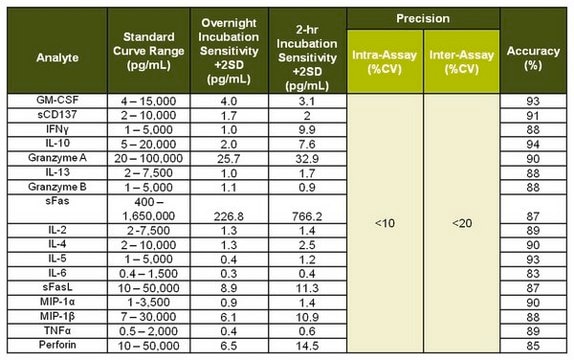
standard curve range: 12.2-50,000 pg/mL
(IL-6)
standard curve range: 2.4-10,000 pg/mL
(Granzyme B, IL-4, TNFα)
standard curve range: 24.4-100,000 pg/mL
(sFas, sFas Ligand, GM-CSF)
standard curve range: 3.7-15,000 pg/mL
(IL-5)
standard curve range: 4.9-20,000 pg/mL
(sCD137 IFNγ, IL-2, MIP-1ß, RANTES)
standard curve range: 48.8-200,000 pg/mL
(IL-13)
standard curve range: 6.1-25,000 pg/mL
(IL-10)
technique(s)
multiplexing: suitable
detection method
fluorometric (Luminex xMAP)
shipped in
wet ice
Related Categories
General description
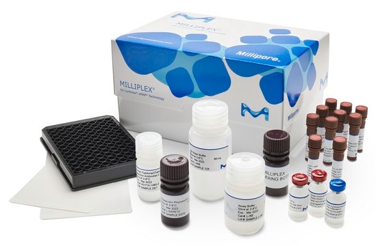
The MILLIPLEX™ Mouse CD8+ T Cell Bead Panel enables you to focus on the therapeutic potential of Mouse CD8+ T Cell analytes. Coupled with the Luminex® xMAP® platform in a bead format, you receive the advantage of ideal speed and sensitivity, allowing quantitative multiplex detection of several analytes simultaneously, which can dramatically improve productivity.
The MILLIPLEX® Mouse CD8+ T Cell Bead Panel is the most versatile system available for Mouse CD8+ T Cell research.
This MILLIPLEX® kit offers you the ability to:
- Select a 15-plex premixed option or
- Choose any combination of analytes from our panel of 15 analytes to design a custom kit that better meets your needs.
- A convenient “all-in-one” box format gives you the assurance that you will have all the necessary reagents you need to run your assay.
Panel Type: Cytokines/Chemokines
Specificity
There was no or minimum cross-reactivity between the antibodies and any other analytes in the panel (see details in assay protocol)..
Application
- Analytes: sCD137, sFas, sFAS ligand, GM-CSF, Granzyme B, IFNγ, IL-2, IL-4, IL-5, IL-6, IL-10, IL-13, MIP-1β, RANTES, TNF-α
- Recommended Sample type: serum, plasma or tissue/cell lysate and culture supernatant
- Recommended Sample dilution: neat
- Assay Run Time: One day or overnight
- Research Category: Inflammation & Immunology
Features and Benefits
Storage and Stability
Other Notes
Legal Information
Disclaimer
signalword
Warning
Hazard Classifications
Acute Tox. 4 Dermal - Acute Tox. 4 Inhalation - Acute Tox. 4 Oral - Aquatic Chronic 2 - Eye Irrit. 2 - Skin Irrit. 2 - Skin Sens. 1 - STOT SE 3
target_organs
Respiratory system
wgk_germany
WGK 3
Certificates of Analysis (COA)
Search for Certificates of Analysis (COA) by entering the products Lot/Batch Number. Lot and Batch Numbers can be found on a product’s label following the words ‘Lot’ or ‘Batch’.
Already Own This Product?
Find documentation for the products that you have recently purchased in the Document Library.
Related Content
See how multiplexing the inflammation signaling pathway with MILLIPLEX® inflammation assays or cell signaling assays can help researchers bridge the gap between immunology and cell signaling, including investigating T cell signaling, Th Cell differentiation, inflammatory response signaling, and sepsis signaling.
Our team of scientists has experience in all areas of research including Life Science, Material Science, Chemical Synthesis, Chromatography, Analytical and many others.
Contact Technical Service