SCT140
BioTracker 555 UV-Excitation Red Lysosome Dye
Live cell imaging dye for acidic cellular organelles such as lysosomes.
Synonym(s):
Live cell imaging probe
Sign Into View Organizational & Contract Pricing
All Photos(1)
About This Item
UNSPSC Code:
12352207
Recommended Products
technique(s)
cell based assay: suitable
detection method
fluorometric
shipped in
ambient
Related Categories
General description
Lysosomes are membrane-enclosed organelles that contain an array of enzymes capable of breaking down all types of biological material including proteins, nucleic acids, carbohydrates, and lipids. Lysosomes function as the digestive system of the cell, serving both to degrade material taken up from outside the cell and to digest obsolete components of the cell itself.
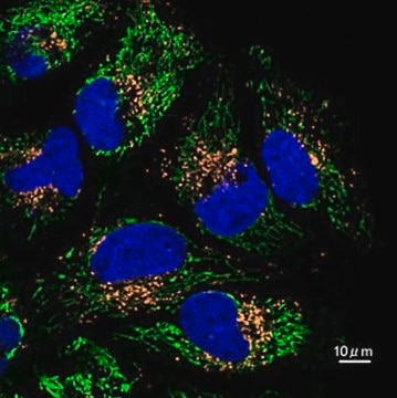
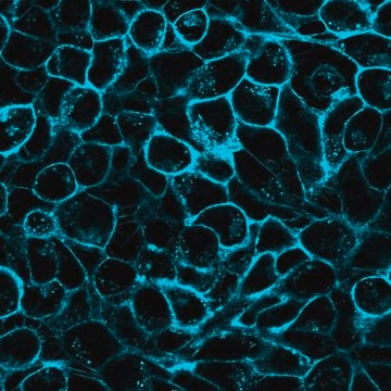
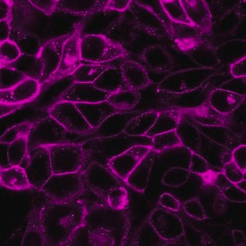
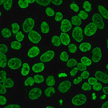
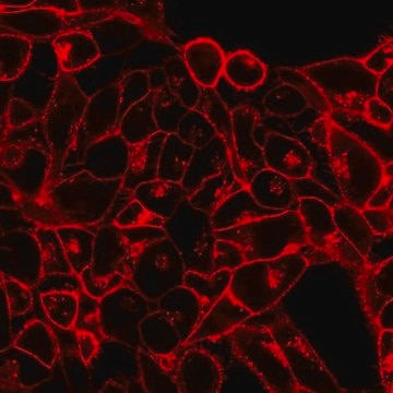
BioTracker Lysosome dyes are fluorescent stains for imaging lysosome localization and morphology in live cells. The dyes accumulate in the low pH environment of the lysosomes, resulting in highly specific lysosomal staining without the need for a wash step.
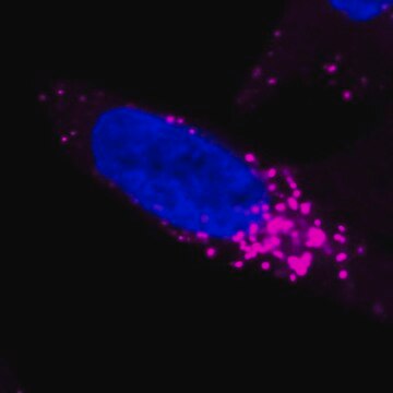
The BioTracker 555 UV-Excitation Red Lysosome is a red fluorogenic lysosome dye with pH-dependent fluorescence. The dye is unique among commercially available lysosome dyes in that its fluorescence in cells is activated by exposure to UV excitation. In solution, the dye shows pH-dependent fluorescence that does not require UV activation. The dye initially shows low fluorescence, but brief exposure to UV excitation from a mercury arc lamp through a DAPI filter.
Spectral Properties
Absorbance: 554nm
Emission: 583nm
Application
Live cell fluorescent imaging
Live cell imaging dye for acidic cellular organelles such as lysosomes.
Research Category
Cell Imaging
Cell Imaging
Research Sub Category
Live Cell Dye
Live Cell Dye
Quality
Spectral Properties
Absorbance: 554nm
Emission: 583nm
Absorbance: 554nm
Emission: 583nm
Physical form
Liquid
Storage and Stability
Store BioTracker 555 UV-Excitation Red Lysosome Dye at -20ºC. Protect From Light.
Note: Centrifuge vial briefly to collect contents at bottom of vial before opening.
Note: Centrifuge vial briefly to collect contents at bottom of vial before opening.
Disclaimer
Unless otherwise stated in our catalog or other company documentation accompanying the product(s), our products are intended for research use only and are not to be used for any other purpose, which includes but is not limited to, unauthorized commercial uses, in vitro diagnostic uses, ex vivo or in vivo therapeutic uses or any type of consumption or application to humans or animals.
Storage Class
10 - Combustible liquids
wgk_germany
WGK 1
flash_point_f
188.6 °F - (refers to pure substance)
flash_point_c
87 °C - (refers to pure substance)
Certificates of Analysis (COA)
Search for Certificates of Analysis (COA) by entering the products Lot/Batch Number. Lot and Batch Numbers can be found on a product’s label following the words ‘Lot’ or ‘Batch’.
Already Own This Product?
Find documentation for the products that you have recently purchased in the Document Library.
Customers Also Viewed
Our team of scientists has experience in all areas of research including Life Science, Material Science, Chemical Synthesis, Chromatography, Analytical and many others.
Contact Technical Service