Recommended Products
form
liquid
Quality Level
usage
sufficient for ≥100 slides staining and fixing
application(s)
diagnostic assay manufacturing
hematology
histology
storage temp.
2-8°C
Related Categories
Application
Silver Enhancer Kit has been used to determine the presence of gold nanorods in various tissues and to prepare silver enhanced gold colloids for electron microscopy.
Features and Benefits
Colloidal gold labels are normally visible only at the electron microscope level. Silver Enhancer enlarges the gold colloid label by precipitation of metallic silver to give a high contrast signal visible under a light microscope. A darkroom is not necessary for this procedure, as the silver enhancer working solution is light-stable for 20-30 minutes under ordinary laboratory lighting.
Packaging
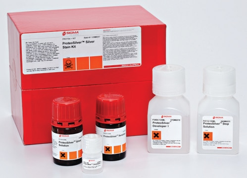
The Silver Enhancer kit consists of Solution A (silver salt), Solution B (initiator), Sodium Thiosulfate (fixer), and a Technical Bulletin describing their use.
Principle
Solution A and Solution B are mixed 1:1 immediately before use and applied to the slide holding the gold-labeled section. After 5-10 minutes, the slide is rinsed with distilled water, fixed for 2-3 minutes in a sodium thiosulfate solution, and rinsed again. The section is now ready for counterstaining and mounting as required.
Kit Components Only
Product No.
Description
- Sodium thiosulfate pentahydrate, reagent grade, ≥99.5% 500 g
signalword
Danger
hcodes
Hazard Classifications
Aquatic Acute 1 - Aquatic Chronic 1 - Repr. 1B
wgk_germany
WGK 3
flash_point_f
Not applicable
flash_point_c
Not applicable
Choose from one of the most recent versions:
Certificates of Analysis (COA)
Lot/Batch Number
Sorry, we don't have COAs for this product available online at this time.
If you need assistance, please contact Customer Support.
Already Own This Product?
Find documentation for the products that you have recently purchased in the Document Library.
Using Immuno-Scanning Electron Microscopy
for the Observation of Focal Adhesion-substratum
interactions at the Nano- and Microscale in S-Phase Cells
for the Observation of Focal Adhesion-substratum
interactions at the Nano- and Microscale in S-Phase Cells
Manus J.P. Biggs
Cell Culture Labfax (2011)
Kathleen Grennan et al.
Talanta, 68(5), 1591-1600 (2008-10-31)
With lower limits of detection and increased stability constantly being demanded of biosensor devices, characterisation of the constituent layers that make up the sensor has become unavoidable, since this is inextricably linked with its performance. This work describe the optimisation
Tsung-Ting Tsai et al.
Scientific reports, 7(1), 3155-3155 (2017-06-11)
In this study, we investigated the antibacterial activity of silver-coated gold nanoparticles (Au-Ag NPs) immobilized on cellulose paper. Ag NPs are known to have strong antibacterial properties, while Au NPs are biocompatible and relatively simple to prepare. We made the
Kumiko A Percival et al.
Cell reports, 41(2), 111484-111484 (2022-10-13)
Midget and parasol ganglion cells (GCs) represent the major output channels from the primate eye to the brain. On-type midget and parasol GCs exhibit a higher background spike rate and thus can respond more linearly to contrast changes than their
A histological evaluation and in vivo assessment of intratumoral near infrared photothermal nanotherapy-induced tumor regression
Hadiyah N
International journal of nanomedicine (2014)
Our team of scientists has experience in all areas of research including Life Science, Material Science, Chemical Synthesis, Chromatography, Analytical and many others.
Contact Technical Service