A3316
Anti-Human IgG (Fc Specific)−Agarose antibody produced in goat
affinity isolated antibody, PBS suspension
Synonym(s):
Goat anti-human IgG
Sign Into View Organizational & Contract Pricing
All Photos(1)
About This Item
Recommended Products
biological source
goat
Quality Level
conjugate
agarose conjugate
antibody form
affinity isolated antibody
antibody product type
secondary antibodies
clone
polyclonal
form
PBS suspension
species reactivity
human
should not react with
rat, mouse
technique(s)
Ouchterlony double diffusion: suitable
capacity
2 mg/mL, resin binding capacity (human IgG)
storage temp.
2-8°C
target post-translational modification
unmodified
Related Categories
General description
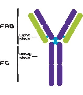
Human IgGs are glycoprotein antibodies that contain two equivalent light chains and a pair of identical heavy chains. IgGs have four distinct isoforms, ranging from IgG1 to IgG4. These antibodies regulate immunological responses to allergy and pathogenic infections. IgGs have also been implicated in complement fixation and autoimmune disorders.
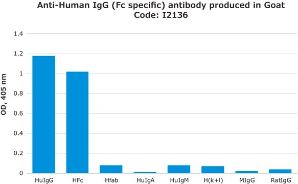
Goat Anti-Human IgG (Fc Specific)-Agarose antibody is specific for the Fc fragment of human IgG when tested against purified human IgA, IgG, IgM, Fc, and kappa and lambda light chains. No reactivity with mouse or rat IgG is seen by Ouchterlony Double Diffusion (ODD), prior to agarose bead coupling.
Goat Anti-Human IgG (Fc Specific)-Agarose antibody is specific for the Fc fragment of human IgG when tested against purified human IgA, IgG, IgM, Fc, and kappa and lambda light chains. No reactivity with mouse or rat IgG is seen by Ouchterlony Double Diffusion (ODD), prior to agarose bead coupling.
Immunogen
Fc fragment of human IgG.
Application
Anti-Human IgG (Fc Specific)-Agarose antibody produced in goat has also been used to purify galanin 1 (Gala1), 3Gal-substituted P-selectin glycoprotein ligand-1 (PSGL-1)/hIgG1 from supernatants of transfected COS cells.
Goat Anti-Human IgG (Fc Specific)-Agarose antibody is suitable for use in Ouchterlony double diffusion. The antibody has also been used for affinity purification assays.
Immunoprecipitation assays were performed using goat anti-human IgG (Fc specific) crosslinked to agarose beads. The beads were diluted in 1 ml of 5% FBS/DMEM for 2 hours at 4 degrees.
Other Notes
Antibody adsorbed with mouse and rat IgG
Physical form
Suspension in 0.01 M phosphate buffered saline, pH 7.4, containing 15 mM sodium azide
Disclaimer
Unless otherwise stated in our catalog or other company documentation accompanying the product(s), our products are intended for research use only and are not to be used for any other purpose, which includes but is not limited to, unauthorized commercial uses, in vitro diagnostic uses, ex vivo or in vivo therapeutic uses or any type of consumption or application to humans or animals.
Not finding the right product?
Try our Product Selector Tool.
wgk_germany
WGK 3
Certificates of Analysis (COA)
Search for Certificates of Analysis (COA) by entering the products Lot/Batch Number. Lot and Batch Numbers can be found on a product’s label following the words ‘Lot’ or ‘Batch’.
Already Own This Product?
Find documentation for the products that you have recently purchased in the Document Library.
Customers Also Viewed
Rebecca J Brown et al.
The Journal of clinical endocrinology and metabolism, 102(6), 1789-1791 (2016-12-03)
Hyperinsulinemia can lead to pathologic ovarian growth and androgen production. A 29-year-old woman developed an autoantibody to the insulin receptor (type B insulin resistance), causing extreme insulin resistance and hyperinsulinemia. Testosterone levels were elevated to the adult male range. Treatment
J L Xu et al.
Immunity, 13(1), 37-45 (2000-08-10)
All rearranging antigen receptor genes have one or two highly diverse complementarity determining regions (CDRs) among the six that typically form the ligand binding surface. We report here that, in the case of antibodies, diversity at one of these regions
J Liu et al.
Transplantation, 63(11), 1673-1682 (1997-06-15)
The hyperacute rejection caused by preformed natural antibodies in the recipient species reacting with donor species endothelial antigens is one of the major obstacles preventing routine use of clinical xenotransplantation. Based on the known structure and biosynthetic pathway of the
S Sumitran et al.
Experimental neurology, 159(2), 347-361 (1999-10-03)
Transplantation of porcine embryonic brain cells, including dopaminergic neurons, from ventral mesencephalon (VM) is considered a potential treatment for patients with Parkinson's disease. In the present study, we characterized the distribution among VM cells of the major porcine endothelial xenoantigen
Jining Liu et al.
Xenotransplantation, 10(2), 149-163 (2003-02-18)
Hyperacute organ xenograft rejection can be prevented by removing anti-pig antibodies by extracorporeal absorption prior to transplantation. A novel recombinant absorber of anti-pig antibodies was developed by fusing the cDNA encoding the extracellular part of a mucin-type protein, P-selectin glycoprotein
Our team of scientists has experience in all areas of research including Life Science, Material Science, Chemical Synthesis, Chromatography, Analytical and many others.
Contact Technical Service