Skip to Content
MilliporeSigma
All Photos(5)

Documents

A4200

Sigma-Aldrich

Monoclonal Anti-γ-Adaptin antibody produced in mouse

clone 100/3, ascites fluid

Synonym(s):

Anti-AP-1

Sign Into View Organizational & Contract Pricing


About This Item

MDL number:
UNSPSC Code:
12352203
NACRES:
NA.41

biological source

mouse

Quality Level

conjugate

unconjugated

antibody form

ascites fluid

antibody product type

primary antibodies

clone

100/3, monoclonal

mol wt

antigen 104 kDa

contains

15 mM sodium azide

species reactivity

bovine, human, monkey

should not react with

mouse, rat

technique(s)

electron microscopy: suitable
immunoprecipitation (IP): suitable
western blot: 1:100 using bovine brain extract

isotype

IgG2b

UniProt accession no.

shipped in

dry ice

storage temp.

−20°C

target post-translational modification

unmodified

Gene Information

human ... AP1G1(164)
mouse ... Ap1g1(11765)
rat ... Ap1g1(171494)

General description

Clathrin-coated vesicle populations contain the adaptor complexes AP-1 and AP-2, also known as HA-I (HA1) adaptor and HA-II (HA2) adaptor or Assemble Protein 1 (AP1) and Assemble Protein 2 (AP2), respectively. Clathrin-coated membranes at or originating from the plasma membrane contain the AP-2 adaptor, while the AP-1 adaptor appears to be largely restricted to clathrin-coated membranes of the trans-Golgi network. Both the Golgi-associated AP-1 adaptor and the plasma-membrane associated AP-2 adaptor are heterotetrameric protein complexes. AP-1 adaptor consists of four subunits termed b1 (formerly b′, 110 kDa), g (100 kDa), m1 (47 kDa), and s1 (20 kDa). b1 and g subunits (b1 and g adaptins) from neuronal sources behave in standard SDS-PAGE like 115 kDa and 104 kDa polypeptides, respectively.

Specificity

Mouse monoclonal clone 100/3 anti-g-Adaptin antibody reacts in immunoblotting with the 104 kDa polypeptide of the Golgi adaptor complex AP-1. It reacts with polypeptides of ~100 kDa in bovine liver, human heart fibroblasts, and Madin-Darby bovine kidney cultured cells (MDBK), but not with any components in the 110-115 kDa range from these sources or from neuroblastoma or astrocytes. This suggests that the 110 and 115 kDa polypeptides may be specific variants occurring only in some cell types of brain. The antibody does not recognize the γ-subunit in rodents (rat and mouse).

Immunogen

AP-1 adaptor from bovine brain.

Application

Applications in which this antibody has been used successfully, and the associated peer-reviewed papers, are given below.
Immuno-electron microscopy (1 paper)
Immunofluorescence (1 paper)
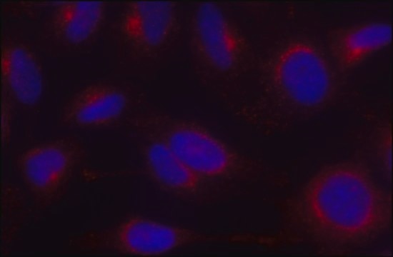
Immunofluorescence of Hela cells was performed using monoclonal anti-AP1 (clone 100/3) as the primary antibody.
In immuno­fluorescence using MDBK cells, human heart fibroblasts, and African green monkey kidney cells (Vero), the binding of antibody appears to be largely confined to the trans-Golgi network. The antibody has been used for studies on the effects of Brefeldin A, a substance causing rapid redistribution of coat proteins associated with the clathrin-coated vesicles that bud from the trans-Golgi network. The antibody is also useful for the immunoaffinity purification of the Golgi adaptor complex AP-1.
Mouse monoclonal anti-gamma-adaptin antibody can be used for western blot (1:100, using bovine brain extract), immunoprecipitation and electron microscopy applications.

Physical form

The product is provided as ascites fluid with 0.1% sodium azide as a preservative.

Disclaimer

Unless otherwise stated in our catalog or other company documentation accompanying the product(s), our products are intended for research use only and are not to be used for any other purpose, which includes but is not limited to, unauthorized commercial uses, in vitro diagnostic uses, ex vivo or in vivo therapeutic uses or any type of consumption or application to humans or animals.

Not finding the right product?  

Try our Product Selector Tool.

Storage Class

13 - Non Combustible Solids

wgk_germany

WGK 1

flash_point_f

Not applicable

flash_point_c

Not applicable


Certificates of Analysis (COA)

Search for Certificates of Analysis (COA) by entering the products Lot/Batch Number. Lot and Batch Numbers can be found on a product’s label following the words ‘Lot’ or ‘Batch’.

Already Own This Product?

Find documentation for the products that you have recently purchased in the Document Library.

Visit the Document Library

Wiebke Stahlschmidt et al.
The Journal of biological chemistry, 289(8), 4906-4918 (2014-01-11)
Clathrin plays important roles in intracellular membrane traffic including endocytosis of plasma membrane proteins and receptors and protein sorting between the trans-Golgi network (TGN) and endosomes. Whether clathrin serves additional roles in receptor recycling, degradative sorting, or constitutive secretion has
E Conway de Macario et al.
Journal of immunology (Baltimore, Md. : 1950), 129(4), 1670-1674 (1982-10-01)
Sixty-nine hybridomas were generated to produce monoclonal antibodies to species of methanogens representing three of the four families accepted at the present time: Methanobacteriaceae, Methanococcaceae, and Methanomicrobiaceae. The antibody of each of 29 hybridomas cross-reacted with a methanogen of the
Nicole A Ducharme et al.
Cellular logistics, 1(2), 57-68 (2011-06-21)
The Rab11 Family Interacting Proteins (Rab11-FIPs) are hypothesized to regulate sequential steps in the apical recycling and transcytotic pathways of polarized epithelial cells. Previous studies have suggested that Rab11-FIP proteins assemble into multi-protein complexes regulating plasma membrane recycling. Rab11-FIP2 interacts
Brogan Yarzabek et al.
eLife, 7 (2018-07-11)
The highly polymorphic human leukocyte antigen (HLA) class I molecules present peptide antigens to CD8+ T cells, inducing immunity against infections and cancers. Quality control mediated by peptide loading complex (PLC) components is expected to ensure the cell surface expression
Jorge Cancino et al.
Molecular biology of the cell, 18(12), 4872-4884 (2007-09-21)
The epithelial-specific adaptor AP1B sorts basolateral plasma membrane (PM) proteins in both biosynthetic and recycling routes, but the site where it carries out this function remains incompletely defined. Here, we have investigated this topic in Fischer rat thyroid (FRT) epithelial

Our team of scientists has experience in all areas of research including Life Science, Material Science, Chemical Synthesis, Chromatography, Analytical and many others.

Contact Technical Service