General description
Monoclonal Anti-VSV-G-Peroxidase is a lyophilized preparation of the purified immunoglobulin fraction of monoclonal Anti-VSV-G (mouse IgG1 isotype) isolated from ascites fluid of the P5D4 hybridoma, conjugated to horseradish peroxidase (HRP). The antibody is derived from the P5D4 hybridoma produced by the fusion of mouse myeloma cells and splenocytes from BALB/c mouse immunized with a synthetic peptide. Vesicular Stomatitis Virus Glycoprotein (VSV-G) exists as a trimer and comprises of four domains, among which domain III is a pleckstrin homology (PH) domain.
Specificity
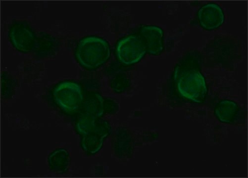
The antibody recognizes an epitope containing the five carboxy-terminal amino acids of VSV Glycoprotein. In infected cells, the antibody localizes the immature forms of VSV-G in the rough endoplasmic reticulum (RER) and in the cisternae of Golgi complex, as well as mature VSV-G at the cell surface and in the budding virus. The antibody does not stain the secreted form of VSV-G which lacks the membrane and the cytoplasmic domain. This antibody has been used for studies on the role of the cytoplasmic domain on newly-synthesized VSV-G during transfer to the plasma membrane and cell surface, using micro-injected antibody, immunoblotting, immunoprecipitation, immunocytochemistry and immunoelectron microscopy. The antibody has been used for the detection, immunoprecipitation and immunocytochemical staining of exogenously introduced constructs tagged with the carboxyl-terminus of VSV-G. This tag does not interfere with the function of the studied protein and can be specifically recognized by the P5D4 antibody without cross-reaction with any endogenous protein.
VSV-G tag sequence (YTDIEMNRLGK) on VSV-G tagged fusion proteins when expressed N- or C-terminal to the fusion protein. Recognizes native and denatured forms of VSV-G tagged proteins.
Immunogen
synthetic peptide containing the 15 carboxy-terminal amino acids (497-511) of Vesicular Stomatitis Virus Glycoprotein (VSV-G), conjugated to KLH.
Application
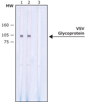
Monoclonal Anti-VSV-G−Peroxidase antibody produced in mouse may be used immunoblotting.
Biochem/physiol Actions
Vesicular Stomatitis Virus Glycoprotein (VSV-G) amino acid sequence YTDIEMNRLGK, corresponding to amino acids 501-511 has been widely used as an epitope tag in expression vectors, enabling the expression of proteins as VSV-G tagged fusion proteins. It constitutes an attractive model to study maturation and intracellular transport of membrane proteins. VSV-G mediates attachment of VSV to the cell surface and induces pH-dependent fusion between viral and target membranes. Besides, its cytoplasmic domain contains information for several intracellular sorting steps, which include efficient export from the endoplasmic reticulum, basolateral delivery and endocytosis..VSV-G is transported through stacks in the Golgi cisternae.
Physical form
Supplied as a lyophilized powder. After reconstitution,the solution contains 1% BSA and 0.05% MIT in 0.01 M sodium phosphate buffered saline, pH 7.4.
Reconstitution
Reconstitution with 0.5 mL water results in a solution of 0.01 M sodium phosphate buffered saline, pH 7.4, containing 1% BSA and 0.05% MIT.
Storage and Stability
Stored the lyophilized product at 2-8 °C. After reconstitution, for extended storage, freeze in working aliquots at –20 °C. For continuous use, the solution may be store at 2-8 °C for up to 1 month. Working dilutions should be discarded. Avoid repeated freeze-thaw.
Disclaimer
Unless otherwise stated in our catalog, our products are intended for research use only and are not to be used for any other purpose, which includes but is not limited to, unauthorized commercial uses, in vitro diagnostic uses, ex vivo or in vivo therapeutic uses or any type of consumption or application to humans or animals.
