CLL1143
SKOV3 Cells GFP-HER2 RFP-EGFR
human female ovary (Source Disease: Ovarian adenocarcinoma)
Sign Into View Organizational & Contract Pricing
All Photos(7)
About This Item
UNSPSC Code:
41106514
NACRES:
NA.81
Recommended Products
product name
SKOV3 Cells GFP-HER2 RFP-EGFR,
biological source
human female ovary (Source Disease: Ovarian adenocarcinoma)
Quality Level
storage temp.
−196°C
Gene Information
human ... EGFR(1956) , ERBB1(1956) , ERBB2(2064) , HER2(2064)
General description
SKOV3 GFP-HER2 RFP-EGFR are ovarian adenocarcinoma cells from a human 64 year old caucasion female having two ZFN modifications - a HER2-GFP transgene and a EGFR-RFP transgene both expressed from their corresponding endogenous gene locus.
This cell line was derived from ATCC Catalog No. HTB-77.
This cell line was derived from ATCC Catalog No. HTB-77.
Application
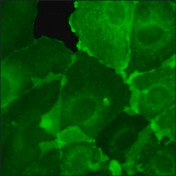
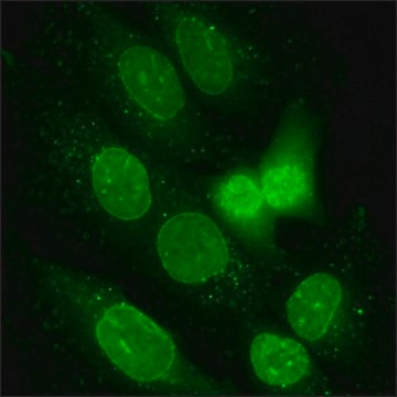
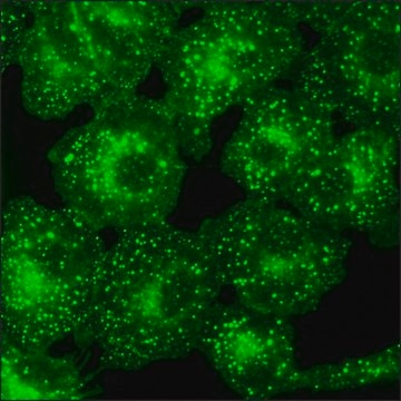
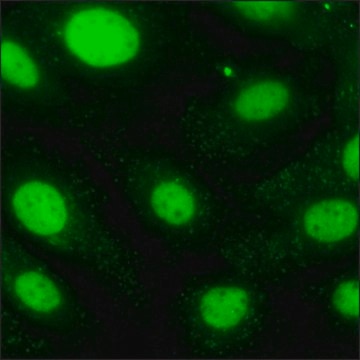
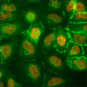
This product is a human SKOV3 cell line in which the genomic HER2 gene has been endogenously tagged with a Green Fluorescent Protein (GFP) gene and the genomic EGFR gene with a Red Fluorescent Protein (RFP) gene using CompoZr® Zinc Finger Nuclease technology. Integration resulted in endogenous expression of a fusion protein in which GFP is attached to the C-terminus of HER2 and of a fusion protein in which RFP is attached to the C-terminus of EGFR. Flourescence imaging shows chacteristic HER2 and EGFR membrane expression. Upon the addition of EGF, the cell line shows redistribution of EGFR from the cell membrane to endosomes, making it useful for high content screening of compounds. For example, the redistribution can be abolished by a selective inhibitor of EGFR, Tyrphostin AG 1478. This stable cell line was expanded from a single clone. The target′s gene regulation and corresponding protein function are preserved in contrast to cell lines with overexpression via an exogenous promoter.
Features and Benefits
Zinc Finger Nuclease (ZFN)-mediated targeted integrations of a fluorescent GFP tag into the last coding exon of the HER2 gene on chromosome 17q21.1 and an RFP tag into the last coding exon of the EGFR gene on chromosome 7p11.2 to create a cell line exhibiting stable expression of both transgenes - each tagged with the corresponding fluorescent reporter on the C-terminus of the protein.
These SKOV3 cells are adherent with a doubling time of approx. 48 hours.
These SKOV3 cells are adherent with a doubling time of approx. 48 hours.
Quality
Tested for Mycoplasma, sterility, post-freeze viability, short terminal repeat (STR) analysis for cell line identification, cytochrome oxidase I (COI) analysis for cell line species confirmation.
Preparation Note
Media Renewal changes two to three times per week.
Rapidly thaw vial by gentle agitation in 37°C water bath (~2 minutes), keeping vial cap out of the water. Decontaminate with 70% ethanol, add 9 mL culture media and centrifuge 125 x g (5-7 minutes). Resuspend in complete culture media and incubate at 37°C in a 5% CO2 atmosphere.
Subculture Ratio: approx. 1:3-1:6
The base medium for this cell line is McCoy′s 5A Medium, Cat. No. M8403. To make the complete growth medium, add the following components to the base medium: fetal bovine serum, Cat. No. F2442, to a final concentration (v/v) of 10% and L-glutamine, Cat. No. G7513, at a final concentration of 1.5 mM.
Cell freezing medium-DMSO 1X, Cat. No. C6164.
Legal Information
CompoZr is a registered trademark of Merck KGaA, Darmstadt, Germany
Disclaimer
RESEARCH USE ONLY. This product is regulated in France when intended to be used for scientific purposes, including for import and export activities (Article L 1211-1 paragraph 2 of the Public Health Code). The purchaser (i.e. enduser) is required to obtain an import authorization from the France Ministry of Research referred in the Article L1245-5-1 II. of Public Health Code. By ordering this product, you are confirming that you have obtained the proper import authorization.
wgk_germany
WGK 3
flash_point_f
closed cup
flash_point_c
closed cup
Certificates of Analysis (COA)
Search for Certificates of Analysis (COA) by entering the products Lot/Batch Number. Lot and Batch Numbers can be found on a product’s label following the words ‘Lot’ or ‘Batch’.
Already Own This Product?
Find documentation for the products that you have recently purchased in the Document Library.
Sebastian Strauss et al.
Nature methods, 17(8), 789-791 (2020-07-01)
DNA-PAINT's imaging speed has recently been significantly enhanced by optimized sequence design and buffer conditions. However, this implementation has not reached an ultimate speed limit and is only applicable to imaging of single targets. To further improve acquisition speed, we
Our team of scientists has experience in all areas of research including Life Science, Material Science, Chemical Synthesis, Chromatography, Analytical and many others.
Contact Technical Service