F0552
Fas Ligand from mouse
>95% (SDS-PAGE), recombinant, expressed in mouse NSO cells, lyophilized powder
Sign Into View Organizational & Contract Pricing
All Photos(1)
About This Item
Recommended Products
recombinant
expressed in mouse NSO cells
Quality Level
assay
>95% (SDS-PAGE)
form
lyophilized powder
mol wt
monomer calculated mol wt ~18 kDa
28-32 kDa by SDS-PAGE
impurities
endotoxin, tested
UniProt accession no.
storage temp.
−20°C
Gene Information
mouse ... Fasl(14103)
General description
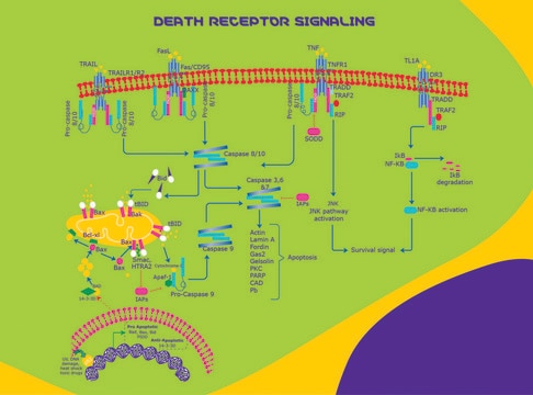
FASLG (Fas ligand) acts as a ligand for Fas receptor, and is a major protein involved in programmed cell death, apoptosis. Soluble Fas (sFAS) is usually detected in plasma prior to apoptosis.
Application
Fas Ligand (FASLG) from mouse has been used for-
- the induction of apoptosis in PC12 cells and
- the induction of migration in BV-2 murine microglial cells.
Biochem/physiol Actions
FASLG (Fas ligand) and Fas receptor constitute the basic elements in apoptosis. Interaction of FASLG with Fas receptor leads to activation of caspase-8. This caspase in turn leads to activation of effector caspases such as caspase-3, -6 and -7. This cascade results in the hydrolysis of nuclear and cytoplasmic components. Expression of FASLG is induced by nuclear factor-κB (NFκB). NFκB/FASLG pathway facilitates the suppression of p,p′-DDT (dichlorodiphenoxytrichloroethane)-induced cell toxicity by vitamin C and E. In CD4+ T cells, this protein is expressed on stimulus by T-cell receptor (TCR), both during normal and pathological conditions, such as alcohol exposure.
Fas ligand, a protein belonging to the tumor necrosis factor (TNF) family of cytokines, induces apoptosis in cells expressing the cell membrane receptor Fas (CD95/Apo-1).
Other Notes
Mouse Fas Ligand, N-terminal 6X histidine-tagged, encodes amino acid residues 132-279.
Physical form
Lyophilized from a 0.2 μm filtered solution in phosphate buffered saline containing 2.5 mg bovine serum albumin.
Analysis Note
Measured by its ability to induce apoptosis in Jurkat cells.
wgk_germany
WGK 3
flash_point_f
Not applicable
flash_point_c
Not applicable
Certificates of Analysis (COA)
Search for Certificates of Analysis (COA) by entering the products Lot/Batch Number. Lot and Batch Numbers can be found on a product’s label following the words ‘Lot’ or ‘Batch’.
Already Own This Product?
Find documentation for the products that you have recently purchased in the Document Library.
Xiaoting Jin et al.
PloS one, 9(12), e113257-e113257 (2014-12-03)
Dichlorodiphenoxytrichloroethane (DDT) is a known persistent organic pollutant and liver damage toxicant. However, there has been little emphasis on the mechanism underlying liver damage toxicity of DDT and the relevant effective inhibitors. Hence, the present study was conducted to explore
Aleksander Szymanowski et al.
Atherosclerosis, 233(2), 616-622 (2014-02-19)
Apoptosis of natural killer (NK) cells is increased in patients with coronary artery disease (CAD) and may explain why NK cell levels are altered in these patients. Soluble forms of Fas and Fas ligand (L) are considered as markers of
Nicole Suyun Liu et al.
PloS one, 7(8), e43180-e43180 (2012-08-21)
Diva is a member of the Bcl2 family but its function in apoptosis remains largely unclear because of its specific expression found within limited adult tissues. Previous overexpression studies done on various cell lines yielded conflicting conclusions pertaining to its
Ying-mei Lu et al.
Journal of neuroinflammation, 9, 172-172 (2012-07-14)
The cerebral microvascular occlusion elicits microvascular injury which mimics the different degrees of stroke severity observed in patients, but the mechanisms underlying these embolic injuries are far from understood. The Fas ligand (FasL)-Fas system has been implicated in a number
Smita S Ghare et al.
Journal of immunology (Baltimore, Md. : 1950), 193(1), 412-421 (2014-06-06)
Activation-induced Fas ligand (FasL) mRNA expression in CD4+ T cells is mainly controlled at transcriptional initiation. To elucidate the epigenetic mechanisms regulating physiologic and pathologic FasL transcription, TCR stimulation-responsive promoter histone modifications in normal and alcohol-exposed primary human CD4+ T
Our team of scientists has experience in all areas of research including Life Science, Material Science, Chemical Synthesis, Chromatography, Analytical and many others.
Contact Technical Service