Skip to Content
MilliporeSigma
All Photos(3)

Documents

HPA004128

Sigma-Aldrich

Anti-PLP1 antibody produced in rabbit

enhanced validation

Prestige Antibodies® Powered by Atlas Antibodies, affinity isolated antibody, buffered aqueous glycerol solution

Synonym(s):

Anti-GPM6C, Anti-PLP, Anti-SPG2

Sign Into View Organizational & Contract Pricing


About This Item

UNSPSC Code:
12352203
Human Protein Atlas Number:
NACRES:
NA.41

biological source

rabbit

Quality Level

conjugate

unconjugated

antibody form

affinity isolated antibody

antibody product type

primary antibodies

clone

polyclonal

product line

Prestige Antibodies® Powered by Atlas Antibodies

form

buffered aqueous glycerol solution

species reactivity

human

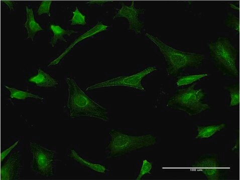
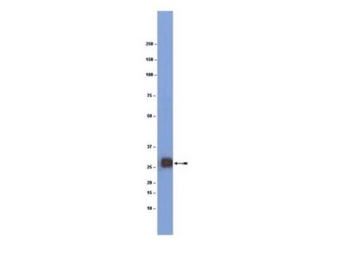
enhanced validation

orthogonal RNAseq
Learn more about Antibody Enhanced Validation

technique(s)

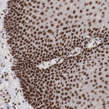
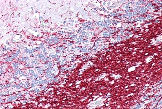
immunohistochemistry: 1:500-1:1000

immunogen sequence

LLLAEGFYTTGAVRQIFGDYKTTICGKGLSATVTGGQKGRGSRGQHQAHSLERVCHCLGKWLGHPDKFVGI

UniProt accession no.

shipped in

wet ice

storage temp.

−20°C

target post-translational modification

unmodified

Gene Information

human ... PLP1(5354)

General description

Proteolipid protein 1 (PLP1) gene encodes for two major proteins of the central nervous system (CNS) myelin, such as proteolipid protein (PLP) and DM20. This gene is located on the human chromosome at Xq22.2.

Immunogen

Myelin proteolipid protein recombinant protein epitope signature tag (PrEST)

Biochem/physiol Actions

Proteolipid protein (PLP) is involved in membrane adhesion, compaction of myelin and synthesis of myelin intra-period line. It is also involved in the maturation of oligodendrocytes. PLP plays a role in wrapping, maintenance and survival of axons. Mutations in the proteolipid protein 1 (PLP1) gene is associated with Pelizaeus-Merzbacher disease indicated by nystagmus, spasticity, microcephaly, ataxia and intellectual disability. PLP1 gene plays a role in Xq22.2 microdeletion and microduplication syndromes. Duplication of this gene results in the overexpression of PLP, which leads to accumulation in the cytoplasm myelinating oligodendrocytes and defects in myelin.

Features and Benefits

Prestige Antibodies® are highly characterized and extensively validated antibodies with the added benefit of all available characterization data for each target being accessible via the Human Protein Atlas portal linked just below the product name at the top of this page. The uniqueness and low cross-reactivity of the Prestige Antibodies® to other proteins are due to a thorough selection of antigen regions, affinity purification, and stringent selection. Prestige antigen controls are available for every corresponding Prestige Antibody and can be found in the linkage section.

Every Prestige Antibody is tested in the following ways:
  • IHC tissue array of 44 normal human tissues and 20 of the most common cancer type tissues.
  • Protein array of 364 human recombinant protein fragments.

Linkage

Corresponding Antigen APREST74534

Physical form

Solution in phosphate-buffered saline, pH 7.2, containing 40% glycerol and 0.02% sodium azide

Legal Information

Prestige Antibodies is a registered trademark of Merck KGaA, Darmstadt, Germany

Disclaimer

Unless otherwise stated in our catalog or other company documentation accompanying the product(s), our products are intended for research use only and are not to be used for any other purpose, which includes but is not limited to, unauthorized commercial uses, in vitro diagnostic uses, ex vivo or in vivo therapeutic uses or any type of consumption or application to humans or animals.

Not finding the right product?  

Try our Product Selector Tool.

wgk_germany

WGK 1

flash_point_f

Not applicable

flash_point_c

Not applicable

ppe

Eyeshields, Gloves, multi-purpose combination respirator cartridge (US)


Certificates of Analysis (COA)

Search for Certificates of Analysis (COA) by entering the products Lot/Batch Number. Lot and Batch Numbers can be found on a product’s label following the words ‘Lot’ or ‘Batch’.

Already Own This Product?

Find documentation for the products that you have recently purchased in the Document Library.

Visit the Document Library

Ravinder Pannu et al.
Journal of neuroscience research, 79(3), 340-350 (2004-12-18)
Spinal cord injury (SCI) is a devastating and complex clinical condition involving proinflammatory cytokines and nitric oxide toxicity that produces a predictable pattern of progressive injury entailing neuronal loss, axonal destruction, and demyelination at the site of impact. The involvement
Derek P Ng et al.
Biochemistry, 53(23), 3747-3757 (2014-05-27)
Central to the formation of tertiary structure in membrane protein folding is the presence of amino acid sequence motifs (such as "small-XXX-small" segments) in the TM segments that promote interaction-compatible surfaces through which the TM α-helices interact. Here, we sought
P Martínez-Montero et al.
Clinical genetics, 84(6), 566-571 (2013-01-26)
Pelizaeus-Merzbacher disease (PMD) is caused in most cases by either duplications or point mutations in the PLP1 gene. This disease, a dysmyelinating disorder affecting mainly the central nervous system, has a wide clinical spectrum and its causing mutations act through
Dorota Hoffman-Zacharska et al.
Medycyna wieku rozwojowego, 17(4), 293-300 (2014-02-13)
The Pelizaeus-Merzbacher disease (PMD) is a rare X-linked recessive hypomyelination disorder caused by mutations of the proteolipid protein1 gene (PLP1). There is a spectrum of PLP1-related disorders from very severe connatal PMD, through classical PMD to mild spastic paraplegia type
A microdeletion at Xq22. 2 implicates a glycine receptor GLRA4 involved in intellectual disability, behavioral problems and craniofacial anomalies
Labonne J D J, et al.
BMC Neurology, 16(1), 1-12 (2016)

Our team of scientists has experience in all areas of research including Life Science, Material Science, Chemical Synthesis, Chromatography, Analytical and many others.

Contact Technical Service