Skip to Content
MilliporeSigma
All Photos(7)

Documents

HPA006462

Sigma-Aldrich

Anti-S100A1 antibody produced in rabbit

enhanced validation

Prestige Antibodies® Powered by Atlas Antibodies, affinity isolated antibody, buffered aqueous glycerol solution

Synonym(s):

Anti-Protein S100-A1 antibody produced in rabbit, Anti-S-100 protein α-chain antibody produced in rabbit, Anti-S-100 protein α-subunit antibody produced in rabbit, Anti-S100 calcium-binding protein A1 antibody produced in rabbit

Sign Into View Organizational & Contract Pricing


About This Item

UNSPSC Code:
12352203
Human Protein Atlas Number:
NACRES:
NA.41

biological source

rabbit

Quality Level

conjugate

unconjugated

antibody form

affinity isolated antibody

antibody product type

primary antibodies

clone

polyclonal

product line

Prestige Antibodies® Powered by Atlas Antibodies

form

buffered aqueous glycerol solution

species reactivity

human

enhanced validation

orthogonal RNAseq
recombinant expression
Learn more about Antibody Enhanced Validation

technique(s)

immunoblotting: 0.04-0.4 μg/mL
immunohistochemistry: 1:500-1:1000

immunogen sequence

GSELETAMETLINVFHAHSGKEGDKYKLSKKELKELLQTELSGFLDAQKDVDAVDKVMKELDENGDGEVDFQEYVVLVAALTVACNNFFWENS

UniProt accession no.

application(s)

research pathology

shipped in

wet ice

storage temp.

−20°C

target post-translational modification

unmodified

Gene Information

human ... S100A1(6271)

Immunogen

Protein S100-A1 recombinant protein epitope signature tag (PrEST)

Application

Applications in which this antibody has been used successfully, and the associated peer-reviewed papers, are given below.
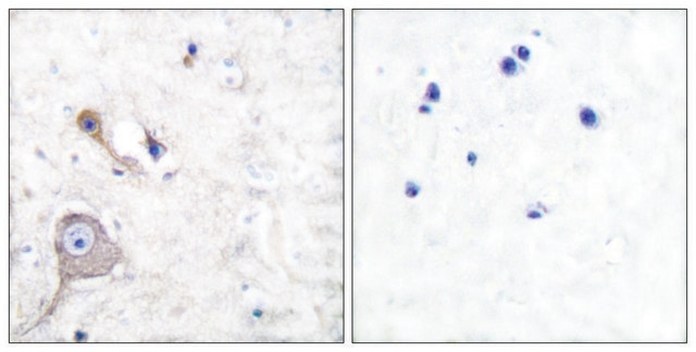
Immunohistochemistry (1 paper)

Biochem/physiol Actions

S100 calcium-binding protein A1 (S100A1) is a protein encoded by the S100A1 gene in humans. It is referred to as S100, S100A and S100-α and belongs to Ca2+-binding S100 protein family. The protein is a homodimer of noncovalently bound subunits. It is a multifunctional regulatory protein involved in a variety of biological processes and closely associated with several human diseases. This gene is expressed in brain and heart tissue, where it plays an important role as a modulator of Ca2+ homeostasis, energy metabolism, neurotransmitter release and contractile performer. Abnormal expression of this protein is associated with neurodegenerative and inflammatory disorders, myopathies and cancer.

Features and Benefits

Prestige Antibodies® are highly characterized and extensively validated antibodies with the added benefit of all available characterization data for each target being accessible via the Human Protein Atlas portal linked just below the product name at the top of this page. The uniqueness and low cross-reactivity of the Prestige Antibodies® to other proteins are due to a thorough selection of antigen regions, affinity purification, and stringent selection. Prestige antigen controls are available for every corresponding Prestige Antibody and can be found in the linkage section.

Every Prestige Antibody is tested in the following ways:
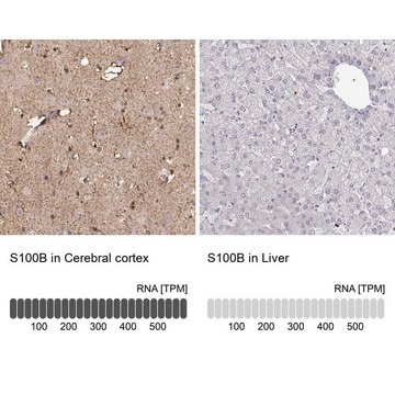
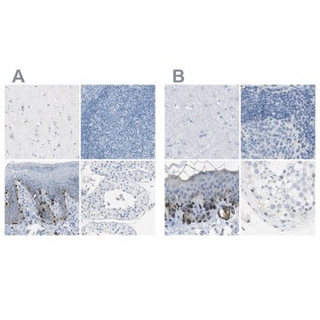
  • IHC tissue array of 44 normal human tissues and 20 of the most common cancer type tissues.
  • Protein array of 364 human recombinant protein fragments.

Linkage

Corresponding Antigen APREST70718

Physical form

Solution in phosphate-buffered saline, pH 7.2, containing 40% glycerol and 0.02% sodium azide

Legal Information

Prestige Antibodies is a registered trademark of Merck KGaA, Darmstadt, Germany

Disclaimer

Unless otherwise stated in our catalog or other company documentation accompanying the product(s), our products are intended for research use only and are not to be used for any other purpose, which includes but is not limited to, unauthorized commercial uses, in vitro diagnostic uses, ex vivo or in vivo therapeutic uses or any type of consumption or application to humans or animals.

Not finding the right product?  

Try our Product Selector Tool.

wgk_germany

WGK 1

flash_point_f

Not applicable

flash_point_c

Not applicable

ppe

Eyeshields, Gloves, multi-purpose combination respirator cartridge (US)


Certificates of Analysis (COA)

Search for Certificates of Analysis (COA) by entering the products Lot/Batch Number. Lot and Batch Numbers can be found on a product’s label following the words ‘Lot’ or ‘Batch’.

Already Own This Product?

Find documentation for the products that you have recently purchased in the Document Library.

Visit the Document Library

Keng Lim Ng et al.
Translational andrology and urology, 8(Suppl 2), S123-S137 (2019-06-27)
Differentiation of chromophobe renal cell carcinoma (chRCC) from benign renal oncocytoma (RO) can be challenging especially when there are overlapping histological and morphological features. In this study we have investigated immunohistochemical biomarkers (cytokeratin 7/CK7, Caveolin-1/Cav-1 and S100 calcium-binding protein A1/S100A1)
Anastasia Malek et al.
Clinical & experimental metastasis, 27(4), 261-271 (2010-04-07)
Tumor cell dissemination to distant organs accounts for the majority of cancer related deaths. Analysis of the stepwise process of metastasis formation and progression might provide novel therapeutic strategies for the treatment of disseminated cancer. However, studies with both biological
Michał Nowakowski et al.
Journal of structural biology, 174(2), 391-399 (2011-02-08)
S100A1 belongs to the EF-hand superfamily of calcium binding proteins. It is a representative of the S100 protein family based on amino acid sequence, three-dimensional structure, and biological function as a calcium signal transmitter. It is a homodimer of noncovalently
Michał Nowakowski et al.
Biochemistry, 52(7), 1149-1159 (2013-01-29)
S100 proteins play a crucial role in multiple important biological processes in vertebrate organisms acting predominantly as calcium signal transmitters. S100A1 is a typical representative of this family of proteins. After four Ca(2+) ions bind, it undergoes a dramatic conformational
Martina Lenarčič Živković et al.
The Journal of biological chemistry, 287(48), 40457-40470 (2012-09-20)
S100A1 protein is a proposed target of molecule-guided therapy for heart failure. S-Nitrosylation of S100A1 is present in cells, increases Ca(2+) binding, and tunes the overall protein conformation. Thiol-aromatic molecular switch is responsible for NO-related modification of S100A1 properties. Post-translational

Our team of scientists has experience in all areas of research including Life Science, Material Science, Chemical Synthesis, Chromatography, Analytical and many others.

Contact Technical Service