Skip to Content
MilliporeSigma
All Photos(6)

Documents

HPA029005

Sigma-Aldrich

Anti-SRSF6 antibody produced in rabbit

enhanced validation

Prestige Antibodies® Powered by Atlas Antibodies, affinity isolated antibody, buffered aqueous glycerol solution

Synonym(s):

Anti-B52, Anti-SFRS6, Anti-SRP55, Anti-splicing factor, arginine/serine-rich 6

Sign Into View Organizational & Contract Pricing


About This Item

MDL number:
UNSPSC Code:
12352203
Human Protein Atlas Number:
NACRES:
NA.43

biological source

rabbit

Quality Level

conjugate

unconjugated

antibody form

affinity isolated antibody

antibody product type

primary antibodies

clone

polyclonal

product line

Prestige Antibodies® Powered by Atlas Antibodies

form

buffered aqueous glycerol solution

species reactivity

human, mouse

enhanced validation

orthogonal RNAseq
Learn more about Antibody Enhanced Validation

technique(s)

immunoblotting: 0.04-0.4 μg/mL
immunohistochemistry: 1:50-1:200

immunogen sequence

KDEYEKSRSRSRSRSPKENGKGDIKSKSRSRSQSRSNSPLPVPPSKARSVSPPPKRATSRSR

shipped in

wet ice

storage temp.

−20°C

target post-translational modification

unmodified

Gene Information

human ... SFRS6(6431)

General description

SRSF6 (serine/arginine-rich splicing factor 6) is also known as SRp55. It is a member of arginine/serine-rich splicing factors family. It is one of the predominant splicing factors and can be up-regulated by DNA damage without p53.

Immunogen

splicing factor, arginine/serine-rich 6 recombinant protein epitope signature tag (PrEST)

Application

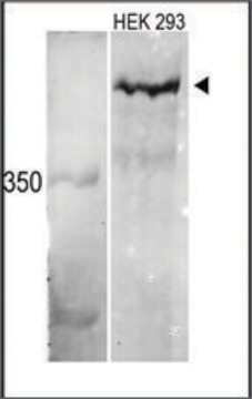
Anti-SRSF6 antibody produced in rabbit has been used in western blotting.

Biochem/physiol Actions

SRSF6 (serine/arginine-rich splicing factor 6) is involved in the generation of BimS (Bcl2-interacting mediator of cell death) mRNA. It also plays a crucial role in Zn2+-induced BimS production. It participates in both constitutive and alternative splicing. It stimulates hyperplasia of sensitized skin. It is a proto-oncogene often expressed in human skin cancer.

Features and Benefits

Prestige Antibodies® are highly characterized and extensively validated antibodies with the added benefit of all available characterization data for each target being accessible via the Human Protein Atlas portal linked just below the product name at the top of this page. The uniqueness and low cross-reactivity of the Prestige Antibodies® to other proteins are due to a thorough selection of antigen regions, affinity purification, and stringent selection. Prestige antigen controls are available for every corresponding Prestige Antibody and can be found in the linkage section.

Every Prestige Antibody is tested in the following ways:
  • IHC tissue array of 44 normal human tissues and 20 of the most common cancer type tissues.
  • Protein array of 364 human recombinant protein fragments.

Linkage

Corresponding Antigen APREST77719

Physical form

Solution in phosphate-buffered saline, pH 7.2, containing 40% glycerol and 0.02% sodium azide.

Legal Information

Prestige Antibodies is a registered trademark of Merck KGaA, Darmstadt, Germany

Disclaimer

Unless otherwise stated in our catalog or other company documentation accompanying the product(s), our products are intended for research use only and are not to be used for any other purpose, which includes but is not limited to, unauthorized commercial uses, in vitro diagnostic uses, ex vivo or in vivo therapeutic uses or any type of consumption or application to humans or animals.

Not finding the right product?  

Try our Product Selector Tool.

wgk_germany

WGK 1

flash_point_f

Not applicable

flash_point_c

Not applicable


Certificates of Analysis (COA)

Search for Certificates of Analysis (COA) by entering the products Lot/Batch Number. Lot and Batch Numbers can be found on a product’s label following the words ‘Lot’ or ‘Batch’.

Already Own This Product?

Find documentation for the products that you have recently purchased in the Document Library.

Visit the Document Library

Comparative expression patterns and diagnostic efficacies of SR splicing factors and HNRNPA1 in gastric and colorectal cancer
Park WC, et al.
BMC Cancer, 16 (2016)
Jorge Rubén Cabrera et al.
Brain pathology (Zurich, Switzerland), 27(2), 181-189 (2016-04-22)
Dendritic alteration of striatal medium spiny neurons is one of the earliest morphological abnormalities in Huntington's disease (HD). The main microtubule-associated protein in dendrites is MAP2. The low-molecular weight isoforms of MAP2 (LMW-MAP2) are the juvenile forms resulting from exclusion
Zizhen Si et al.
Journal of experimental & clinical cancer research : CR, 40(1), 78-78 (2021-02-25)
Long non-coding RNAs (lncRNA) are reported to influence colorectal cancer (CRC) progression. Currently, the functions of the lncRNA ZNF561 antisense RNA 1 (ZNF561-AS1) in CRC are unknown. ZNF561-AS1 and SRSF6 expression in CRC patient samples and CRC cell lines was
Jung-Chun Lin et al.
Scientific reports, 7, 44204-44204 (2017-03-10)
A growing body of studies has demonstrated that dysregulated splicing profiles constitute pivotal mechanisms for carcinogenesis. In this study, we identified discriminative splicing profiles of colorectal cancer (CRC) cells compared to adjacent normal tissues using deep RNA-sequencing (RNA-seq). The RNA-seq
Zinc-induced modulation of SRSF6 activity alters Bim splicing to promote generation of the most potent apoptotic isoform BimS
Hara H, et al.
FEBS Journal, 280(14), 3313-3327 (2013)

Our team of scientists has experience in all areas of research including Life Science, Material Science, Chemical Synthesis, Chromatography, Analytical and many others.

Contact Technical Service