MBD0029
Porphyromonas gingivalis FISH probe - Cy3
Probe for fluorescence in situ hybridization (FISH),20 μM in water
Synonym(s):
In-situ hybridization, bacterial fish probes, fish analysis, fish probes, fish technology, fish testing, fluorescence in situ hybridization, fluorescent in situ hybridization, in situ hybridization
About This Item
Recommended Products
Quality Level
technique(s)
FISH: suitable
fluorescence
λex 550 nm; λem 570 nm (Cy3)
shipped in
dry ice
storage temp.
−20°C
General description
sequence in whole and intact cells.1 Microbial FISH allows the visualization, identification and isolation of bacteria due to recognition of ribosomal RNA also in unculturable samples.2
FISH technique can serve as a powerful tool in the microbiome research field by allowing the observation of native microbial populations in diverse microbiome environments, such as samples from human origin (blood3 and tissue4), microbial ecology (solid biofilms5 and aquatic systems6) and plants7. It is strongly recommended to include positive and negative controls in FISH assays to ensure specific binding of the probe of interest and appropriate protocol conditions. We offer positive (MBD0032/33) and negative control (MBD0034/35) probes, that accompany the specific probe of interest.
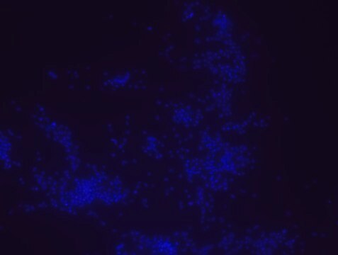
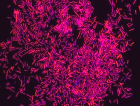
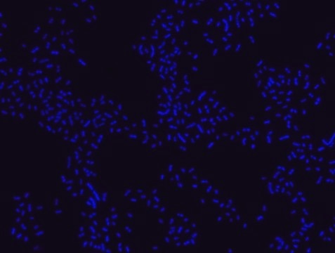
Porphyromonas gingivalis probe specifically recognizes P. gingivalis cells. P. gingivalis, is a gram negative bacterium which is an etiologic agent of adult periodontitis, a chronic inflammatory disease characterized by the destruction of the supportive tissue surrounding teeth. Studies have shown that LPS from P. gingivalis plays an important role in this disease.8-11 The association of the oral microbiota, including P. gingivalis, with various pathological states has been reported. These include development of Alzheimer′s disease12, role in oral cancers13, preterm birth14 and rheumatoid arthritis15. FISH technique was successfully used to identify P. gingivalis with the probe in various samples such as pure culture (as described in the figure legends), dental implants16,17 , periapical tooth lesions18, saliva19, brain tissue20, gingival and aortic tissues21, biofilms from voice prostheses22, subgingival biofilm23, aortic wall tissue24, and infected HeLa cells25. Moreover, FISH can be implicated to identify P. gingivalis in tumor tissue26, multispecies biofilm27, multispecies oral biofilms28 and pure culture and buccal epithelial cells29.
Application
Features and Benefits
- Visualize, identify and isolate Porphyromonas gingivalis cells.
- Observe native P. gingivalis cell populations in diverse microbiome environments.
- Specific, sensitive and robust identification of P. gingivalis in bacterial mixed population.
- Specific, sensitive and robust identification even when P. gingivalis is in low abundance in the sample.
- FISH can complete PCR based detection methods by avoiding contaminant bacteria detection.
- Provides information on P. gingivalis morphology and allows to study biofilm architecture.
- Identify P. gingivalis in clinical samples such as, tumor and brain tissues (for example in formalin-fixed paraffin-embedded (FFPE) samples), saliva and oral cavity and medical equipment such as, dental implants and voice prostheses.
- The ability to detect P. gingivalis in its natural habitat is an essential tool for studying host-microbiome interaction.
Storage Class
12 - Non Combustible Liquids
wgk_germany
nwg
flash_point_f
Not applicable
flash_point_c
Not applicable
Certificates of Analysis (COA)
Search for Certificates of Analysis (COA) by entering the products Lot/Batch Number. Lot and Batch Numbers can be found on a product’s label following the words ‘Lot’ or ‘Batch’.
Already Own This Product?
Find documentation for the products that you have recently purchased in the Document Library.
Our team of scientists has experience in all areas of research including Life Science, Material Science, Chemical Synthesis, Chromatography, Analytical and many others.
Contact Technical Service
