Skip to Content
MilliporeSigma
All Photos(3)

Documents

SAB1400126

Sigma-Aldrich

Anti-HLA-E antibody produced in mouse

IgG fraction of antiserum, buffered aqueous solution

Synonym(s):

Anti-DKFZp686P19218, Anti-EA1.2, Anti-EA2.1, Anti-HLA-6.2, Anti-MHC

Sign Into View Organizational & Contract Pricing


About This Item

UNSPSC Code:
12352203
NACRES:
NA.41

biological source

mouse

Quality Level

conjugate

unconjugated

antibody form

IgG fraction of antiserum

antibody product type

primary antibodies

clone

polyclonal

form

buffered aqueous solution

species reactivity

human

technique(s)

flow cytometry: suitable
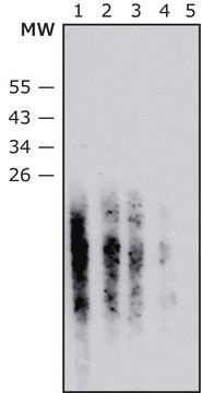
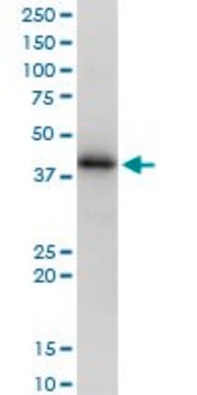
western blot: 1 μg/mL

UniProt accession no.

shipped in

dry ice

storage temp.

−20°C

target post-translational modification

unmodified

Gene Information

human ... HLA-E(3133)

Related Categories

General description

HLA-E belongs to the HLA class I heavy chain paralogues. This class I molecule is a heterodimer consisting of a heavy chain and a light chain (beta-2 microglobulin). The heavy chain is anchored in the membrane. HLA-E binds a restricted subset of peptides derived from the leader peptides of other class I molecules. The heavy chain is approximately 45 kDa and its gene contains 8 exons. Exon one encodes the leader peptide, exons 2 and 3 encode the alpha1 and alpha2 domains, which both bind the peptide, exon 4 encodes the alpha3 domain, exon 5 encodes the transmembrane region, and exons 6 and 7 encode the cytoplasmic tail. (provided by RefSeq)

Immunogen

HLA-E (AAH02578.1, 1 a.a. ~ 358 a.a) full-length human protein.

Sequence
MVDGTLLLLLSEALALTQTWAGSHSLKYFHTSVSRPGRGEPRFISVGYVDDTQFVRFDNDAASPRMVPRAPWMEQEGSEYWDRETRSARDTAQIFRVNLRTLRGYYNQSEAGSHTLQWMHGCELGPDRRFLRGYEQFAYDGKDYLTLNEDLRSWTAVDTAAQISEQKSNDASEAEHQRAYLEDTCVEWLHKYLEKGKETLLHLEPPKTHVTHHPISDHEATLRCWALGFYPAEITLTWQQDGEGHTQDTELVETRPAGDGTFQKWAAVVVPSGEEQRYTCHVQHEGLPEPVTLRWKPASQPTIPIVGIIAGLVLLGSVVSGAVVAAVIWRKKSSGGKGGSYSKAEWSDSAQGSESHSL

Physical form

Solution in phosphate buffered saline, pH 7.4

Not finding the right product?  

Try our Product Selector Tool.

flash_point_f

Not applicable

flash_point_c

Not applicable


Certificates of Analysis (COA)

Search for Certificates of Analysis (COA) by entering the products Lot/Batch Number. Lot and Batch Numbers can be found on a product’s label following the words ‘Lot’ or ‘Batch’.

Already Own This Product?

Find documentation for the products that you have recently purchased in the Document Library.

Visit the Document Library

Damien M Callahan et al.
Journal of applied physiology (Bethesda, Md. : 1985), 116(12), 1582-1592 (2014-05-03)
Age-related loss of skeletal muscle mass and function is implicated in the development of disease and physical disability. However, little is known about how age affects skeletal muscle structure at the cellular and ultrastructural levels or how such alterations impact
Jia Qi et al.
Biological & pharmaceutical bulletin, 37(11), 1713-1718 (2014-11-05)
Heat shock protein 90 (HSP90) is a ubiquitous molecular chaperone involved in the proper conformation of many proteins. HSP90 inhibitors (17-dimethyl aminoethylamino-17-demethoxygeldanamycin hydrochloride [17-DMAG]) bind to and inactivate HSP90, suppressing some key signaling pathways involved in the inflammatory process. Since
Lasse Gliemann et al.
American journal of physiology. Heart and circulatory physiology, 307(8), H1111-H1119 (2014-08-17)
In animal studies, the polyphenol resveratrol has been shown to influence several pathways of importance for angiogenesis in skeletal muscle. The aim of the present study was to examine the angiogenic effect of resveratrol supplementation with parallel exercise training in
Mari Narumi et al.
PloS one, 9(10), e109859-e109859 (2014-10-10)
Compared with the peripheral corneal limbus, the human central cornea lacks blood vessels, which is responsible for its immunologically privileged status and high transparency. Dendritic cells (DCs) are present in the central avascular area of inflamed corneas, but the mechanisms
Laleh Talebian et al.
Transfusion, 54(6), 1515-1521 (2014-01-23)
The NKG2D receptor, one of the natural killer (NK) cell-activating receptors, is expressed on the surface of CD3+CD8+ T cells, γδ+ T cells, NK cells, NKT cells, and a few CD4+ T cells. We show, for the first time, a

Our team of scientists has experience in all areas of research including Life Science, Material Science, Chemical Synthesis, Chromatography, Analytical and many others.

Contact Technical Service