Skip to Content
MilliporeSigma
All Photos(2)

Documents

SAB4200705

Sigma-Aldrich

Anti-Neurofilament 200 (Phos and Non-Phos) antibody, Mouse monoclonal

clone N52, purified from hybridoma cell culture

Synonym(s):

Anti-CMT2CC, Anti-NFH

Sign Into View Organizational & Contract Pricing


About This Item

UNSPSC Code:
12352203
NACRES:
NA.41

antibody form

purified from hybridoma cell culture

Quality Level

antibody product type

primary antibodies

clone

N52, monoclonal

mol wt

~200 kDa

species reactivity

mouse, pig, feline, rat, monkey, bovine, human

concentration

~1.0 mg/mL

technique(s)

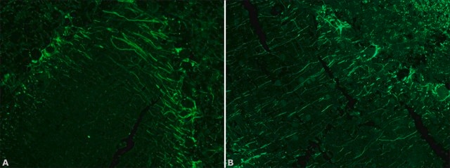
immunoblotting: 2.5-5 μg/mL using human neuroblastoma SH-SY5Y cell line fresh lysate
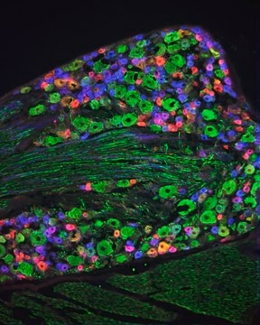
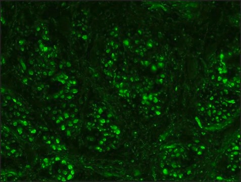
immunofluorescence: suitable
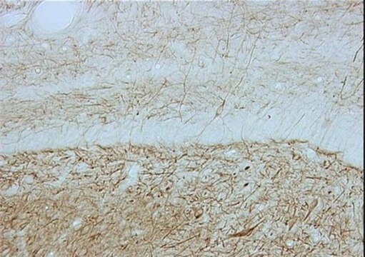
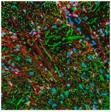
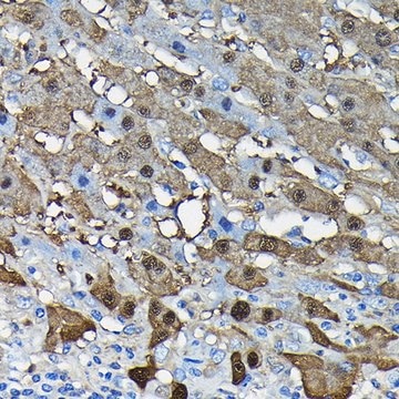
immunohistochemistry: 10 μg/mL using heat-retrieved formalin-fixed, paraffin-embedded human Cerebellum sections

isotype

IgG1

UniProt accession no.

shipped in

dry ice

storage temp.

−20°C

target post-translational modification

unmodified

Gene Information

General description

Monoclonal Anti-Neurofilament 200 (phosphorylated and non-phosphorylated) (mouse IgG1 isotype) is derived from the N52 hybridoma produced by the fusion of mouse myeloma cells and splenocytes from an immunized mouse. Neurofilament 200 also known as Neurofilament heavy polypeptide (H-subunit) and is 200 kDa.
Neurofilament 200 is encoded by the gene located at human chromosome 22q12.2.

Specificity

Monoclonal Anti-Neurofilament 200 (Phos. and NonPhos.) also known as Neurofilament-H or Heavy subunit, specifically recognizes an epitope on the C-terminal tail domain of Neurofilament 200, which is present on both the phosphorylated and nonphosphorylated forms of this glycoprotein. The antibody shows reactivity with neurofilaments in the central and peripheral nervous systems from human , pig, mouse, rat, monkey, feline and bovine origin.

Application

Anti-Neurofilament 200 (Phos and Non-Phos) antibody has been used in
  • immunoblotting
  • immunofluorescence
  • immunohistochemistry

Biochem/physiol Actions

Mutation in the NEFH gene leads to the development of autosomal dominant axonal Charcot-Marie-Tooth disease (CMT2cc).
Neurofilament 200 has an important function in axonal transport, axonal plasticity and maintaining the structure of neuronal cytoskeleton. Defects in Neurofilament 200 are a cause of susceptibility to amyotrophic lateral sclerosis (ALS) and these accumulations are a hallmark of pathological lesion.

Physical form

Solution in 0.01 M phosphate buffered saline pH 7.4, containing 15 mM sodium azide as a preservative.

Storage and Stability

For continuous use, store at 2-8°C for up to one month. For extended storage, freeze in working aliquots. Repeated freezing and thawing is not recommended. If slight turbidity occurs upon prolonged storage, clarify the solution by centrifugation before use. Working dilution samples should be discarded if not used within 12 hours.

Disclaimer

Unless otherwise stated in our catalog or other company documentation accompanying the product(s), our products are intended for research use only and are not to be used for any other purpose, which includes but is not limited to, unauthorized commercial uses, in vitro diagnostic uses, ex vivo or in vivo therapeutic uses or any type of consumption or application to humans or animals.

Still not finding the right product?  

Give our Product Selector Tool a try.

Storage Class

10 - Combustible liquids

flash_point_f

Not applicable

flash_point_c

Not applicable


Certificates of Analysis (COA)

Search for Certificates of Analysis (COA) by entering the products Lot/Batch Number. Lot and Batch Numbers can be found on a product’s label following the words ‘Lot’ or ‘Batch’.

Already Own This Product?

Find documentation for the products that you have recently purchased in the Document Library.

Visit the Document Library

Kenji Kashiwagi et al.
Investigative ophthalmology & visual science, 44(1), 154-159 (2002-12-31)
To investigate the phosphorylation of the heavy neurofilament subunit (NF-H), which could be deeply involved in axonal transport of retinal ganglion cells (RGCs), in an experimental glaucoma model of chronic elevation of intraocular pressure (IOP) in monkeys. One eye in
Dissociation of axonal neurofilament content from its transport rate
Yuan A, et al.
Testing, 10(7), e0133848-e0133848 (2015)
Alterations of a 200 kDa neurofilament in the rat hippocampus after forebrain ischemia
Kaku Y, et al.
Journal of Cerebral Blood Flow and Metabolism, 13(3), 402-408 (1993)
Harry B M Uylings et al.
Psychiatry research, 183(1), 1-20 (2010-06-12)
The orbitofrontal cortex (OFC) is located on the basal surface of the frontal lobe and is distinguished by its unique anatomical and functional features. Clinical and postmortem studies suggest the involvement of the orbitofrontal cortex in psychiatric disorders. However, the
Y-L Liu et al.
Spinal cord, 47(2), 166-170 (2008-07-30)
Observational cross-section study. The objective of our study was to determine if phosphorylation of aggregated neurofilaments (NFs) would occur in autoimmune-mediated motor neuron injury. Our main hypothesis was that autoimmune-mediated damage of spinal cord motor neurons may influence NF phosphorylation

Our team of scientists has experience in all areas of research including Life Science, Material Science, Chemical Synthesis, Chromatography, Analytical and many others.

Contact Technical Service