General description
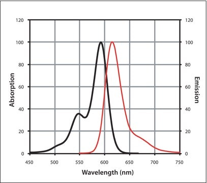
Antibody CF™ 647 conjugates are affinity-purified antibodies labeled with a far-red fluorescent dye, CF647. CF™ 647 conjugates are useful as secondary antibodies for detection.
Immunoglobulin G (IgG) is a major class of immunoglobulin family. It contains gamma (γ) heavy chain in the constant (C) region and is a largely expressed serum antibody. Monomeric structure of IgG contains two identical heavy chains and two identical light chains with molecular weight of 50kDa and 25kDa respectively. The primary structure of this antibody also contains disulfide bonds involved in linking the two heavy chains, linking the heavy and light chains and resides inside the chains. IgG has four sub classes namely, IgG1, IgG2, IgG3, and IgG4 with different heavy chains, named γ1,γ2, γ3, and γ4, respectively. IgG is produced and released from CD5+B cells, the predominant lymphocytes in the neonatal cell repertoire. Maternal IgG is the only antibody transported across the placenta to the fetus. Maternal IgG passively immunizes the infants.
Specificity
Binds all rabbit IgGs
Immunogen
rabbit IgG (H+L)
Application
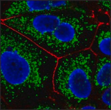
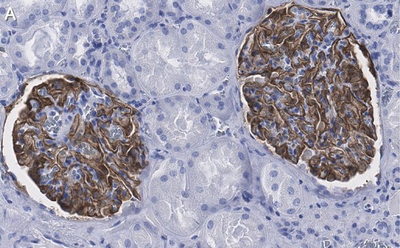
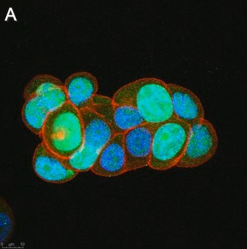
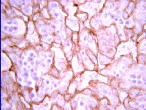
Anti-Rabbit IgG (H+L), CF™ 647 antibody produced in goat has been used as a secondary antibody in sections post terminal deoxynucleotidyl transferase dUTP nick end labeling (TUNEL) analysis of apoptotic cells. It has also been used in immunohistochemical staining at a dilution of 1/1,000.
Features and Benefits
Evaluate our antibodies with complete peace of mind. If the antibody does not perform in your application, we will issue a full credit or replacement antibody.
Learn more.Physical form
Supplied in phosphate buffered saline with 0.05% sodium azide, 50% glycerol and 2 mg/mL bovine serum albumin.
Preparation Note
Protect from light.
Legal Information
This product is distributed by Sigma-Aldrich Co. under the authorization of Biotium, Inc. This product is covered by one or more US patents and corresponding patent claims outside the US patents or pending applications owned or licensed by Biotium, Inc. including without limitation: 12/334,387; 12/607,915; 12/699,778; 12/850,578; 61/454,484. In consideration of the purchase price paid by the buyer, the buyer is hereby granted a limited, non-exclusive, non-transferable license to use only the purchased amount of the product solely for the buyer′s own internal research in a manner consistent with the accompanying product literature. Except as expressly granted herein, the sale of this product does not grant to or convey upon the buyer any license, expressly, by implication or estoppel, under any patent right or other intellectual property right of Biotium, Inc. Buyer shall not resell or transfer this product to any third party, or use the product for any commercial purposes, including without limitation, any diagnostic, therapeutic or prophylactic uses. This product is for research use only. Any other uses, including diagnostic uses, require a separate license from Biotium, Inc. For information on purchasing a license to use this product for purposes other than research, contact Biotium, Inc., 3159 Corporate Place, Hayward, CA 94545, Tel: (510) 265-1027. Fax: (510) 265-1352. Email: btinfo@biotium.com.
CF is a trademark of Biotium, Inc.
Disclaimer
Unless otherwise stated in our catalog or other company documentation accompanying the product(s), our products are intended for research use only and are not to be used for any other purpose, which includes but is not limited to, unauthorized commercial uses, in vitro diagnostic uses, ex vivo or in vivo therapeutic uses or any type of consumption or application to humans or animals.

