05-515
Anti-acetyl-Lysine Antibody, clone 4G12
clone 4G12, Upstate®, from mouse
Sign Into View Organizational & Contract Pricing
All Photos(3)
About This Item
UNSPSC Code:
12352203
eCl@ss:
32160702
NACRES:
NA.41
Recommended Products
biological source
mouse
Quality Level
antibody form
purified immunoglobulin
antibody product type
primary antibodies
clone
4G12, monoclonal
species reactivity
human, mouse, rat, vertebrates
manufacturer/tradename
Upstate®
technique(s)
immunoprecipitation (IP): suitable
western blot: suitable
isotype
IgG
shipped in
dry ice
target post-translational modification
unmodified
General description
In the nucleus, DNA is tightly packed into nucleosomes generating an environment which is highly repressive towards DNA processes such as transcription. Acetylation of lysine residues within proteins has emerged as an important mechanism used by cells to overcome this repression. The acetylation of non-histone proteins such as transcription factors, as well as histones appears to be involved in this process. Acetylation may result in structural transitions as well as specific signaling within discrete chromatin domains. The role of acetylation in intracellular signaling has been inferred from the binding of acetylated peptides by the conserved bromodomain. Furthermore, recent findings suggest that bromodomain/acetylated-lysine recognition can serve as a regulatory mechanism in protein-protein interactions in numerous cellular processes such as chromatin remodeling and transcriptional activation.
Specificity
Acetylated lysine-containing proteins including histones. Of all the possible H3 and H4 acetylation sites, this antibody exhibits highest affinity for histone H4 acetylated on lysine 8 and histone H3 acetylated on lysine 14.
Expected to cross-react with rat and mouse.
Immunogen
Mixture of chemically acetylated antigens; recognizes numerous proteins acetylated on a lysine, most prominently on histones.
Application
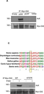
Detect acetyl-Lysine with Anti-acetyl-Lysine Antibody, clone 4G12 (Mouse Monoclonal Antibody), that has been shown to work in IP & WB.
Immunoprecipitation:
5 μg of a previous lot immunoprecipitated in vitro acetylated PCAF added to a 3T3 RIPA cell lysate. The immunoprecipitated PCAF was detected by subsequent western blot analysis using 1 μg/mL monoclonal anti-GST (Catalog # 05-311).
5 μg of a previous lot immunoprecipitated in vitro acetylated PCAF added to a 3T3 RIPA cell lysate. The immunoprecipitated PCAF was detected by subsequent western blot analysis using 1 μg/mL monoclonal anti-GST (Catalog # 05-311).
Quality
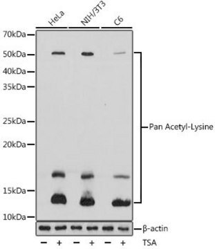
Routinely evaluated by Western Blot on untreated and sodium butyrated treated HeLa lysates.
Western Blot Analysis:
1:500 dilution of this lot detected ACETYL-LYSINE on 10 μg of sodium butyrated treated HeLa lysates.
Western Blot Analysis:
1:500 dilution of this lot detected ACETYL-LYSINE on 10 μg of sodium butyrated treated HeLa lysates.
Target description
Varies depending upon the protein being detected.
Physical form
Format: Purified
Purified mouse monoclonal IgG in buffer containing 0.1 M Tris-glycine, pH 7.4, 0.15 M NaCl, 0.05% sodium azide, before the addition of glycerol to 30%.
Storage and Stability
Stable for 1 year at -20°C from date of receipt. Handling Recommendations: Upon receipt, and prior to removing the cap, centrifuge the vial and gently mix the solution. Aliquot into microcentrifuge tubes and store at -20°C. Avoid repeated freeze/thaw cycles, which may damage IgG and affect product performance. Note: Variability in freezer temperatures below -20°C may cause glycerol containing solutions to become frozen during storage.
Other Notes
Concentration: Please refer to the Certificate of Analysis for the lot-specific concentration.
Legal Information
UPSTATE is a registered trademark of Merck KGaA, Darmstadt, Germany
Not finding the right product?
Try our Product Selector Tool.
wgk_germany
WGK 1
Certificates of Analysis (COA)
Search for Certificates of Analysis (COA) by entering the products Lot/Batch Number. Lot and Batch Numbers can be found on a product’s label following the words ‘Lot’ or ‘Batch’.
Already Own This Product?
Find documentation for the products that you have recently purchased in the Document Library.
Adam W Beharry et al.
Journal of cell science, 127(Pt 7), 1441-1453 (2014-01-28)
The Forkhead box O (FoxO) transcription factors are activated, and necessary for the muscle atrophy, in several pathophysiological conditions, including muscle disuse and cancer cachexia. However, the mechanisms that lead to FoxO activation are not well defined. Recent data from
Wai Chi Ho et al.
Cancer research, 65(10), 4273-4281 (2005-05-19)
The primary goal of chemotherapy is to cause cancer cell death. However, a side effect of many commonly used chemotherapeutic drugs is the activation of nuclear factor-kappaB (NF-kappaB), a potent inducer of antiapoptotic genes, which may blunt the therapeutic efficacy
Contrasting proteome biology and functional heterogeneity of the 20 S proteasome complexes in mammalian tissues.
Gomes, AV; Young, GW; Wang, Y; Zong, C; Eghbali, M; Drews, O; Lu, H; Stefani, E; Ping, P
Molecular and Cellular Proteomics null
Liming Wu et al.
The Journal of biological chemistry, 286(3), 2236-2244 (2010-10-21)
NIR (novel INHAT repressor) is a transcriptional co-repressor with inhibitor of histone acetyltransferase (INHAT) activity and has previously been shown to physically interact with and suppress p53 transcriptional activity and function. However, the mechanism by which NIR suppresses p53 is
FKBP25, a novel regulator of the p53 pathway, induces the degradation of MDM2 and activation of p53.
Anna Maria Ochocka et al.
FEBS letters, 583(4), 621-626 (2009-01-27)
The p53 tumour suppressor protein is tightly controlled by the E3 ubiquitin ligase, mouse double minute 2 (MDM2), but maintains MDM2 expression as part of a negative feedback loop. We have identified the immunophilin, 25kDa FK506-binding protein (FKBP25), previously shown
Our team of scientists has experience in all areas of research including Life Science, Material Science, Chemical Synthesis, Chromatography, Analytical and many others.
Contact Technical Service