General description
Cadherin-5 (UniProt: P33151; also known as 7B4 antigen, Vascular endothelial cadherin, VE-cadherin, CD144) is encoded by the CDH5 gene (Gene ID: 1003) in human. Cadherins are calcium-dependent cell adhesion proteins, which preferentially interact with themselves in a homophilic manner in connecting cells. Cadherin 5 is a single-pass type I membrane protein that is found at cell-cell boundaries and may also be located at cell-matrix boundaries. It is well expressed in brain and endothelial tissue. It plays an important role in endothelial cell biology through control of the cohesion and organization of the intercellular junctions. It associates with alpha-catenin forming a link to the cytoskeleton. Cadherin 5 acts in concert with KRIT1 to establish and maintain correct endothelial cell polarity and vascular lumen. These effects are mediated by recruitment and activation of the Par polarity complex and RAP1B. Cadherin 5 is synthesized as a preproprotein with a signal peptide (aa 1-25) and propeptide (aa 26-47) that are proteolytically cleaved to produce the mature glycoprotein. It has an extracellular domain (aa 48-599), a short transmembrane domain (aa 600-620), and a cytoplasmic domain (aa 621-784) and contains five cadherin domains. Three calcium ions are usually bound at the interface of each cadherin domain and rigidify the connections, providing a strong curvature to the full-length ectodomains. Two isoforms of Cadherin 5 have been described that are produced by alternative splicing. Cadherin 5 can be phosphorylated on tyrosine residues by KDR/VRGFR-2. Phosphorylation at Tyr731 leads to the separation of beta-catenin from the cytoplasmic tail Cadherin 5. This phosphorylation event reported to be sufficient to maintain cells in a mesenchymal state during the cell invasion phase of angiogenesis.
Specificity
This rabbit polyclonal antibody detects human and murine Cadherin-5 phosphorylated on Tyrosine 731.
Immunogen
Epitope: C-terminus
KLH-conjugated linear peptide corresponding to 11 amino acids surrounding phosphorylated tyrosine 731 from human Cadherin-5.
Application
Anti-phospho-VE-cadherin (Tyr731), Cat. No. AB1965-I, is a rabbit polyclonal antibody that detects Cadherin-5 phosphorylated on Tyrosine 731 and has been tested for use in Western Blotting and Peptide Inhibition Assay.
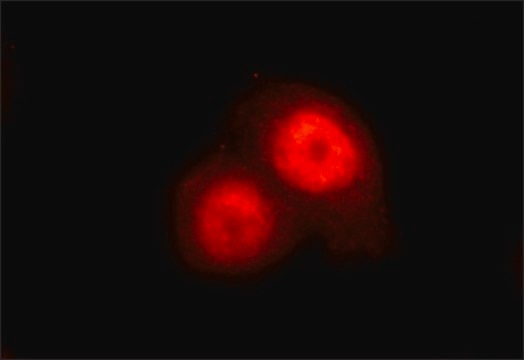
Peptide Inhibition Analysis: A 1:1,500 dilution from a representative lot was used with HUVEC stimulated with VEGF for peptide block analysis.
Research Category
Cell Structure
Quality
Evaluated by Western Blotting in lysate from HUVEC treated with VEGF.
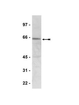
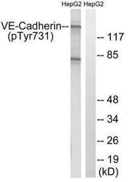
Western Blotting Analysis: A 1:500 dilution of this antibody detected phospho-VE-cadherin (Tyr731) in lysate from HUVEC treated with VEGF.
Target description
~100 kDa observed; 87.53 kDa calculated. Uncharacterized bands may be observed in some lysate(s).
Physical form
Affinity Purified
Purified rabbit polyclonal antibody in buffer containing 0.1 M Tris-Glycine (pH 7.4), 150 mM NaCl with 0.05% sodium azide.
Storage and Stability
Stable for 1 year at 2-8°C from date of receipt.
Other Notes
Concentration: Please refer to lot specific datasheet.
Disclaimer
Unless otherwise stated in our catalog or other company documentation accompanying the product(s), our products are intended for research use only and are not to be used for any other purpose, which includes but is not limited to, unauthorized commercial uses, in vitro diagnostic uses, ex vivo or in vivo therapeutic uses or any type of consumption or application to humans or animals.

