Skip to Content
MilliporeSigma
All Photos(2)

Documents

AB2971

Sigma-Aldrich

Anti-LAMP-1 (CD107a) Antibody

from rabbit, purified by affinity chromatography

Synonym(s):

lysosomal-associated membrane protein 1, lysosome-associated membrane glycoprotein 1, Lysosome-associated membrane protein 1, CD107 antigen-like family member A, CD107a antigen

Sign Into View Organizational & Contract Pricing


About This Item

UNSPSC Code:
12352203
eCl@ss:
32160702
NACRES:
NA.41

biological source

rabbit

Quality Level

antibody form

affinity isolated antibody

antibody product type

primary antibodies

clone

polyclonal

purified by

affinity chromatography

species reactivity

human, mouse, rat

species reactivity (predicted by homology)

rhesus macaque (based on 100% sequence homology)

packaging

antibody small pack of 25 μg

technique(s)

immunocytochemistry: suitable
western blot: suitable

NCBI accession no.

UniProt accession no.

shipped in

ambient

storage temp.

2-8°C

target post-translational modification

unmodified

Gene Information

human ... LAMP1(3916)

General description

Lysosome-associated membrane protein 1 (LAMP-1) is a highly glycosolated, single-pass type 1 membrane protein belonging to the lysosome- associated membrane protein family; a group of proteins that make up the majority of the lysosomal membrane and are considered crucial mediators for cellular adhesion by interacting with carbohydrate receptors. LAMP-1 functions to traffic carbohydrate ligands to selectins. It is also thought to play a role in tumor cell metastasis.

Specificity

The antibody recognizes LAMP-1 at the topological domain (Lumenal).

Immunogen

Epitope: Topological domain (Lumenal)
KLH-conjugated linear peptide corresponding to human LAMP-1 at the topological domain (Lumenal).

Application

Research Category
Apoptosis & Cancer
Research Sub Category
Apoptosis - Additional

Tumor Markers
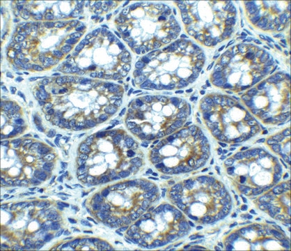
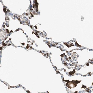
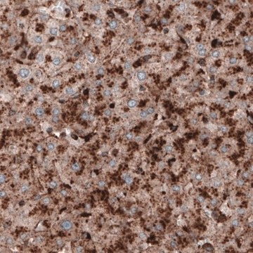
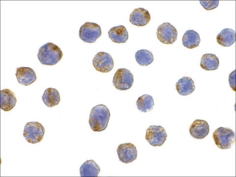
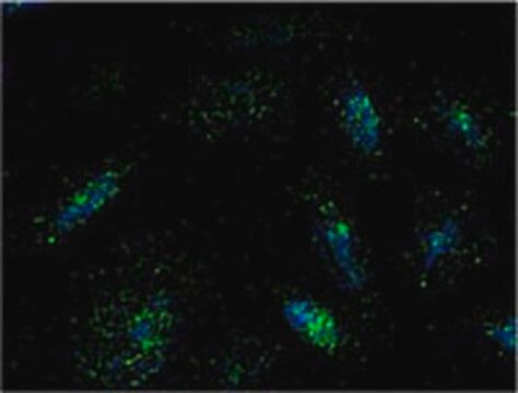
This Anti-LAMP-1 (CD107a) Antibody is validated for use in WB, IC for the detection of LAMP-1 (CD107a).
Western Blot (Snap i.d.) Analysis: 1 µg/mL from a previous lot detected LAMP-1 in 10 µg of EL4 cell lysate.

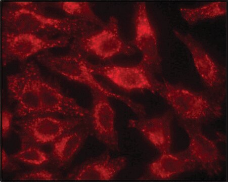
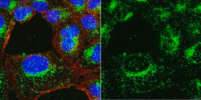
Immunocytochemistry Analysis: 1:500 dilution from a previous lot detected LAMP-1 in NIH/3T3, A431, and HeLa cells.

Quality

Evaluated by Western Blot in EL4 cell lysate.

Western Blot Analysis: 1 µg/mL of this antibody detected LAMP-1 in 10 µg of EL4 cell lysate.

Target description

~ 95kDa observed. Although the calculated MW 45 kDa, LAMP-1 is heavily glycosylated and therefore, MW typically can be expected between 90-120 kDa Uncharacterized bands may be observed in some lysates.

Physical form

Affinity purified
Purified rabbit polyclonal in buffer containing 0.1 M Tris-Glycine (pH 7.4), 150 mM NaCl with 0.05% sodium azide.

Storage and Stability

Stable for 1 year at 2-8°C from date of receipt.

Analysis Note

Control
EL4 cell lysate

Other Notes

Concentration: Please refer to the Certificate of Analysis for the lot-specific concentration.

Disclaimer

Unless otherwise stated in our catalog or other company documentation accompanying the product(s), our products are intended for research use only and are not to be used for any other purpose, which includes but is not limited to, unauthorized commercial uses, in vitro diagnostic uses, ex vivo or in vivo therapeutic uses or any type of consumption or application to humans or animals.

Not finding the right product?  

Try our Product Selector Tool.

wgk_germany

WGK 1

flash_point_f

Not applicable

flash_point_c

Not applicable


Certificates of Analysis (COA)

Search for Certificates of Analysis (COA) by entering the products Lot/Batch Number. Lot and Batch Numbers can be found on a product’s label following the words ‘Lot’ or ‘Batch’.

Already Own This Product?

Find documentation for the products that you have recently purchased in the Document Library.

Visit the Document Library

Carolina Duarte et al.
Journal of cellular and molecular medicine, 26(10), 2841-2851 (2022-04-17)
Emerging studies indicate that intracellular eukaryotic ceramide species directly activate cathepsin B (CatB), a lysosomal-cysteine-protease, in the cytoplasm of osteoclast precursors (OCPs) leading to elevated RANKL-mediated osteoclastogenesis and inflammatory osteolysis. However, the possible impact of CatB on osteoclastogenesis elevated by
Tiago Ferreira et al.
Viruses, 12(10) (2020-10-25)
H-1 protoparvovirus (H-1PV) is a self-propagating virus that is non-pathogenic in humans and has oncolytic and oncosuppressive activities. H-1PV is the first member of the Parvoviridae family to undergo clinical testing as an anticancer agent. Results from clinical trials in
Yoav Charpak-Amikam et al.
Scientific reports, 7(1), 8661-8661 (2017-08-19)
Human cytomegalovirus (HCMV) is a major human pathogen, causing serious diseases in immunocompromised populations and congenially infected neonates. One of the main immune cells acting against the virus are Natural Killer (NK) cells. Killing by NK cells is mediated by
Lucas T Jae et al.
Science (New York, N.Y.), 344(6191), 1506-1510 (2014-06-28)
Lassa virus spreads from a rodent to humans and can lead to lethal hemorrhagic fever. Despite its broad tropism, chicken cells were reported 30 years ago to resist infection. We found that Lassa virus readily engaged its cell-surface receptor α-dystroglycan
Leonardo Ermini et al.
Frontiers in cell and developmental biology, 9, 652651-652651 (2021-05-22)
Aberrant ceramide build-up in preeclampsia, a serious disorder of pregnancy, causes exuberant autophagy-mediated trophoblast cell death. The significance of ceramide accumulation for lysosomal biogenesis in preeclampsia is unknown. Here we report that lysosome formation is markedly increased in trophoblast cells

Articles

Autophagy is a highly regulated process that is involved in cell growth, development, and death. In autophagy cells destroy their own cytoplasmic components in a very systematic manner and recycle them.

Our team of scientists has experience in all areas of research including Life Science, Material Science, Chemical Synthesis, Chromatography, Analytical and many others.

Contact Technical Service