ABE250
Anti-dimethyl Histone H3 (Lys4) Antibody
from rabbit, purified by affinity chromatography
Synonym(s):
H3 histone family, member T, histone 3, H3, histone cluster 3, H3, H3K4me2
About This Item
Recommended Products
biological source
rabbit
Quality Level
antibody form
affinity isolated antibody
antibody product type
primary antibodies
clone
polyclonal
purified by
affinity chromatography
species reactivity
mouse, rat, human, chicken
technique(s)
ChIP: suitable (ChIP-seq)
dot blot: suitable
immunocytochemistry: suitable
western blot: suitable
NCBI accession no.
UniProt accession no.
shipped in
wet ice
target post-translational modification
dimethylation (Lys4)
Gene Information
human ... HIST1H3F(8968)
General description
Specificity
Immunogen
Application
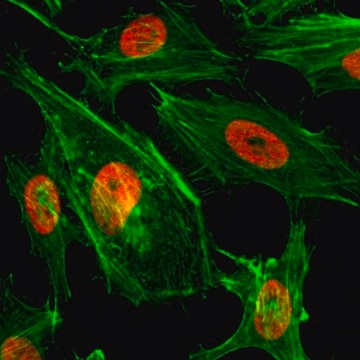
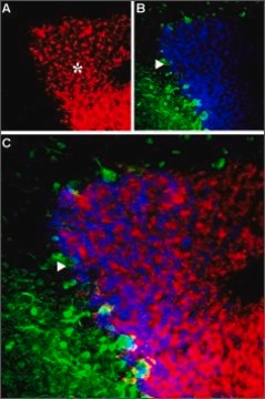
1:500 dilution from a representative lot detected Histone H3 in NIH/3T3 and A431 cells.
Dot Blot Analysis:
1:1,000 dilution from a representative lot specifically detected Histone H3 in a Lys4 dimethhyl modified histone H3 peptide while not detecting potential cross-reacting peptides corresponding to other modified and non-modified histones.
Chromatin Immunoprecipitation Analysis:
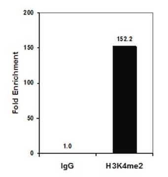
Sonicated chromatin prepared from HeLa cells (1 X 10E6 cell equivalents per IP) were subjected to chromatin immunoprecipitation using 4 µg of either Normal IgG (Part No. 12-370), or 4 µL of Anti-Dimethyl Histone H3 (Lys4) (Part No. ABE250) and the Magna ChIP A/G Kit (Cat. # 17-10085). Successful immunoprecipitation of Dimethyl-Histone H3 (Lys4) associated DNA fragments was verified by qPCR using primers specific for the human GAPDH coding region.
Please refer to the EZ-Magna ChIP A/G (Cat. # 17-10085) protocol for experimental details.
ChIP-Sequencing
Chromatin immunoprecipitation was performed using the Magna ChIP HiSens kit (cat# 17-10460), 5 µg ofa representative lot of anti-dimethyl-Histone H3 (Lys4) antibody(ABE250), 20 µL Protein A/G beads ,and 5e6 crosslinked HeLa cell chromatin followed by DNA purification using magnetic beads. Libraries were prepared from Input and ChIP DNA samples using standard protocols with Illumina barcoded adapters, and analyzed on Illumina HiSeq instrument. An excess of twelve million reads from FastQ files were mapped using Bowtie (http://bowtie-bio.sourceforge.net/manual.shtml) following TagDust (http://genome.gsc.riken.jp/osc/english/dataresource/) tag removal. Peaks were identified using MACS (http://luelab.dfci.harvard.edu/MACS/), with peaks and reads visualized as a custom track in UCSC Genome Browser (http://genome.ucsc.edu) from BigWig and BED files.
Epigenetics & Nuclear Function
Histones
Quality
Western Blot Analysis: 1:1,000 dilution of this antibody detected Histone H3 on 10 µg of HeLa acid extract.
Target description
Linkage
Physical form
Storage and Stability
Analysis Note
HeLa acid extract
Disclaimer
Not finding the right product?
Try our Product Selector Tool.
wgk_germany
WGK 1
flash_point_f
Not applicable
flash_point_c
Not applicable
Certificates of Analysis (COA)
Search for Certificates of Analysis (COA) by entering the products Lot/Batch Number. Lot and Batch Numbers can be found on a product’s label following the words ‘Lot’ or ‘Batch’.
Already Own This Product?
Find documentation for the products that you have recently purchased in the Document Library.
Our team of scientists has experience in all areas of research including Life Science, Material Science, Chemical Synthesis, Chromatography, Analytical and many others.
Contact Technical Service
