Skip to Content
MilliporeSigma
All Photos(1)

Documents

MAB343-C

Sigma-Aldrich

Anti-APP Antibody, CT, clone 2.F2.19B4, Ascites Free

clone 2.F2.19B4, from mouse

Synonym(s):

Amyloid beta A4 protein, ABPP, Alzheimer disease amyloid protein, Amyloid precursor protein, APP, APPI, Cerebral vascular amyloid peptide, CVAP, PN-II, PreA4, Protease nexin-II

Sign Into View Organizational & Contract Pricing


About This Item

UNSPSC Code:
12352203
eCl@ss:
32160702
NACRES:
NA.41

biological source

mouse

Quality Level

antibody form

purified immunoglobulin

antibody product type

primary antibodies

clone

2.F2.19B4, monoclonal

species reactivity

human, mouse

technique(s)

affinity binding assay: suitable
immunocytochemistry: suitable
immunofluorescence: suitable
immunohistochemistry: suitable (paraffin)
immunoprecipitation (IP): suitable
western blot: suitable

isotype

IgG1κ

NCBI accession no.

UniProt accession no.

shipped in

wet ice

target post-translational modification

unmodified

Gene Information

human ... APP(351)

Related Categories

General description

Amyloid beta A4 protein (UniProt P05067; also known as ABPP, Alzheimer disease amyloid protein, Amyloid precursor protein, APP, APPI, Cerebral vascular amyloid peptide, CVAP, PN-II, PreA4, Protease nexin-II) is encoded by the APP (also known as A4, AD1) gene (Gene ID 351) in human. Amyloid precursor protein (APP) is initially produced with a signal peptide sequence (a.a. 1-17), the removal of which yields the mature protein with a large extracellular portion (a.a. 18-699), followed by a transmembrane segment (a.a. 700-723) and a cytoplasmic (a.a. 724-770) tail. APP can be further processed by the α-, β-, and γ-secretases in two alternative processing pathways. In the non-amyloidogenic pathway, APP is first cleaved by the plasma membrane-localized α-secretase to generate an N-terminal extracellular sAPPα fragment (a.a. 18-687) and a membrane-bound C-terminal fragment C83 (CTFα), which can be further cleaved by γ-secretase to produce a non-toxic small peptide p3 and a cytoplasmic APP intracellular domain (AICD). In the amyloidogenic pathway, APP undergoes β-cleavage in BACE-1 (β-site APP-cleaving enzyme)-enriched endosomes to generate an N-terminal extracellular sAPPβ fragment (a.a. 18-671) and a membrane-bound C-terminal fragment C99 (CTFβ). Subsequent cleavage of C99 by γ-secretase releases the amyloid β peptides, Aβ1-42 (672-713) & Aβ1-40 (672-711), and AICD. Aβ accumulation in the cortical and hippocampal regions of the brain is a major pathological feature of Alzheimer′s disease (AD). Aβ Ser8 phosphorylation is shown to promote Aβ aggregation into oligomeric and fibrillar assemblies and to prevent Aβ proteolytic clearance by certain proteases.

Specificity

Clone 2.F2.19B4, also known as the Jonas antibody, regcognizes a C-terminal epitope present in 10 of the 11 human spliced isoforms reported by UniProt (P05067), with APP305 being the only spliced isoform lacking the targeted epitope. Clone 2.F2.19B4 recognizes full-length APP, the APP C-terminal fragments (CTFs) produced by α- and β-secretase cleavage, and the APP intracellular domain (AICD) generated by γ-secretase cleavage, but not the sAPPα or sAPPβ fragment. Epitope has been mapped to APP770 a.a. 732-751, which is equivalent to APP695 a.a. 657-676 (Van Vickle, G.D., et al. (2007). Biochemistry. 46(36):10317-10327).

Immunogen

APP C-terminal 53-a.a. fragment.
Epitope: APP770 a.a. 732-751 (APP695 a.a. 657-676).

Application

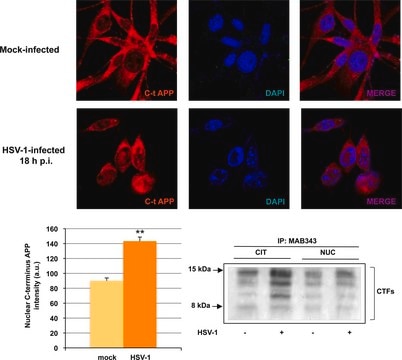
Immunocytochemistry Analysis: A representative lot detected a greatly enhanced nuclear APP immunoreactivity due to an upregulated AICD generation and nuclear localization in HSV-1-infected SH-SY5Y cells by fluorescent immunocytochemistry (De Chiara, G., et al. (2010). PLoS One. 5(11):e13989).
Immunocytochemistry Analysis: A representative lot immunostained neural progenitor cells (NPCs) isolated from the lateral ventricles of E14 mouse brain by fluorescent immunocytochemistry (Ma, Q.H., et al. (2008). Nat. Cell Biol. 10(3):283-294).
Immunoprecipitation Analysis: A representative lot immunoprecipitated APP and cleaved APP C-terminal fragments from uninfected and HSV-1-infected SH-SY5Y cells (De Chiara, G., et al. (2010). PLoS One. 5(11):e13989).
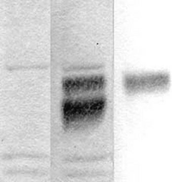
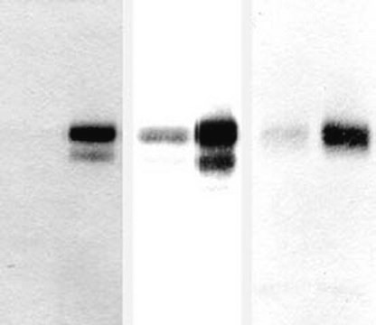
Western Blotting Analysis: A representative lot detected APP and cleaved APP C-terminal fragments in lysates and APP immunoprecipitates from uninfected and HSV-1-infected SH-SY5Y cells (De Chiara, G., et al. (2010). PLoS One. 5(11):e13989).
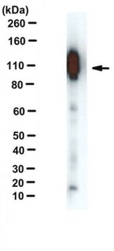
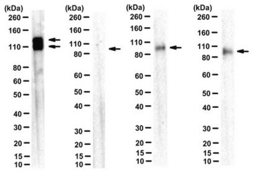
Western Blotting Analysis: Representative lots detected the stably expressed full-length human APP695 in HEK293-derived BA-3 cells (Hwang, E.M., et al. (2008). Bioorg. Med. Chem. 16(14):6669-6674; Yeon, S.W., et al. (2007). Peptides. 28(4):838-844).
Immunofluorescence Analysis: A representative lot immunostained the walls of the lateral ventricles in E14 mouse brain tissue sections by fluorescent immunohistochemistry (Ma, Q.H., et al. (2008). Nat. Cell Biol. 10(3):283-294).
Affinity Binding Assay Analysis: A representative lot captured the synthetic peptide representing APP770 a.a. 732-751 or APP695 a.a. 657-676 (Van Vickle, G.D., et al. (2007). Biochemistry. 46(36):10317-10327).
Research Category
Neuroscience
Research Sub Category
Neurodegenerative Diseases
This Anti-APP Antibody, CT, clone 2.F2.19B4, Ascites Free is validated for use in Immunohistochemistry (Paraffin), Immunocytochemistry, Immunoprecipitation, Western Blotting, Immunofluorescence, Affinity Binding Assay for the detection of APP.

Quality

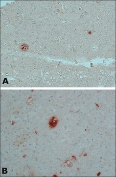
Evaluated by Immunohistochemistry in normal and AD human brain tissue sections.

Immunohistochemistry Analysis: A 1:50 dilution from a representative lot detected APP immunoreactivity in normal human cerebral cortex and Alzheimer′s diseased (AD) brain tissue sections.

Target description

85.18/86.94 kDa (APP770), 75.00/76.76 kDa (L-APP677), 76.90/78.66 kDa (APP695), 77.10/78.87 kDa (L-APP696), 79.01/80.77 kDa (APP714), 81.15/82.92 kDa (L-APP733), 83.06/84.82 kDa (APP751), 83.28/85.04 kDa (L-APP752), 70.79/72.55 kDa (APP639), and 82.76/84.52 kDa (isoform 11) calcualted (full-length mature/pro-form).

Physical form

Format: Purified
Protein G purified.
Purified mouse monoclonal IgG1κ antibody in buffer containing 0.1 M Tris-Glycine (pH 7.4), 150 mM NaCl with 0.05% sodium azide.

Storage and Stability

Stable for 1 year at 2-8°C from date of receipt.

Other Notes

Concentration: Please refer to lot specific datasheet.

Disclaimer

Unless otherwise stated in our catalog or other company documentation accompanying the product(s), our products are intended for research use only and are not to be used for any other purpose, which includes but is not limited to, unauthorized commercial uses, in vitro diagnostic uses, ex vivo or in vivo therapeutic uses or any type of consumption or application to humans or animals.

Not finding the right product?  

Try our Product Selector Tool.

wgk_germany

WGK 1

flash_point_f

Not applicable

flash_point_c

Not applicable


Certificates of Analysis (COA)

Search for Certificates of Analysis (COA) by entering the products Lot/Batch Number. Lot and Batch Numbers can be found on a product’s label following the words ‘Lot’ or ‘Batch’.

Already Own This Product?

Find documentation for the products that you have recently purchased in the Document Library.

Visit the Document Library

Our team of scientists has experience in all areas of research including Life Science, Material Science, Chemical Synthesis, Chromatography, Analytical and many others.

Contact Technical Service