Skip to Content
MilliporeSigma
All Photos(1)

Documents

MABF3036

Sigma-Aldrich

Anti-PSGL-1/CD162 Antibody, clone HECA-452

Synonym(s):

P-selectin glycoprotein ligand 1;PSGL-1 Selectin P ligand,;CD162

Sign Into View Organizational & Contract Pricing


About This Item

UNSPSC Code:
12352203
NACRES:
NA.43

biological source

rat

Quality Level

antibody form

purified antibody

antibody product type

primary antibodies

clone

HECA-452, monoclonal

mol wt

calculated mol wt 43.2 kDa

purified by

affinity chromatography

species reactivity

human

packaging

antibody small pack of 100 μg

technique(s)

flow cytometry: suitable
immunohistochemistry: suitable
western blot: suitable

isotype

IgMκ

epitope sequence

Extracellular domain

Protein ID accession no.

UniProt accession no.

storage temp.

-10 to -25°C

target post-translational modification

unmodified

Gene Information

human ... SELPLG(6404)

Related Categories

General description

P-selectin glycoprotein ligand 1 (UniProt: Q14242; also known as PSGL-1 Selectin P ligand, CD162) is encoded by the SELPLG gene (Gene ID: 6404) in human. Leukocytes express L-selectin ligands that are critical for leukocyte-leukocyte interactions at sites of inflammation. PSGL-1 is a predominant leukocyte L-selectin ligand that displays appropriate sialyl Lewis x (sLex)-like carbohydrate determinants for L-selectin recognition. It is a single-pass, mucin-like type I membrane glycoprotein that is synthesized with a signal peptide (aa 1-17), which is subsequently cleaved off to produce a form that contains an extracellular domain (18-320), a transmembrane domain (321-341), and a cytoplasmic domain (342-412). The mature form is generated following the cleaved of the propeptide (aa 18-41) region. PSGL-1 is a SLe(x)-type proteoglycan, which through high affinity, calcium-dependent interactions with E-, P- and L-selectins mediates rapid rolling of leukocytes over vascular surfaces during the initial steps in inflammation. It is constitutively expressed on all human peripheral-blood T cells and is also expressed on skin-homing T cells. Clone HECA-452 can detect a human endothelial cell differentiation antigen (HECA) selectively expressed on high endothelium. In addition to reacting with high endothelium, it also shows some cross-reactivity with a set of monocytic cells. This clone is shown to block the majority of P-selectin binding to neutrophils. (Ref.: Tu, L., et al. (1999). J. Immunol. 163(9); 5070-5078; Fuhlbrigge, RC., et al. (1997). Nature. 389(6654); 978-981; Duivestijn, AM., et al. (1988). Am. J. Pathol. 130(1); 147-155).

Specificity

Clone HECA-452 is a rat monoclonal antibody that detects PSGL-1/CD162. It targets an epitope within the extracellular domain.

Immunogen

Stromal preparation from human tonsil tissue.

Application

Quality Control Testing

Evaluated by Flow Cytometry in Human peripheral blood mononuclear cells (PBMC).

Flow Cytometry Analysis (FC): 1.0 μg of this antibody stained one million Human peripheral blood mononuclear cells (PBMC).

Tested Applications

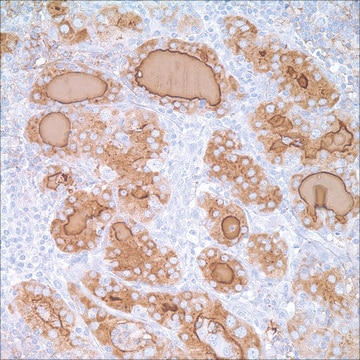
Immunohistochemistry Applications: A representative lot detected PSGL-1/CD162 in Immunohistochemistry application (Duijvestijn, A.M., et al. (1988). Am J Pathol. 130(1):147-55).

Flow Cytometry Analysis: A representative lot detected PSGL-1/CD162 in Flow Cytometry application (Picker, L.J., et al. (1991). Nature. 349(6312):796-9; Fuhlbrigge, R.C., et al. (1997). Nature. 389(6654):978-81).

Western Blotting Analysis: A representative lot detected PSGL-1/CD162 in Western Blotting application (Fuhlbrigge, R.C., et al. (1997). Nature. 389(6654):978-81).

Note: Actual optimal working dilutions must be determined by end user as specimens, and experimental conditions may vary with the end user.

Physical form

Purified rat monoclonal antibody IgM in PBS without azide.

Reconstitution

1.0 mg/mL. Please refer to guidance on suggested starting dilutions and/or titers per application and sample type.

Storage and Stability

Store at -10°C to -25°C. Handling Recommendations: Upon receipt and prior to removing the cap, centrifuge the vial and gently mix the solution. Aliquot into microcentrifuge tubes and store at -20°C. Avoid repeated freeze/thaw cycles, which may damage IgG and affect product performance.

Other Notes

Concentration: Please refer to the Certificate of Analysis for the lot-specific concentration.

Disclaimer

Unless otherwise stated in our catalog or other company documentation accompanying the product(s), our products are intended for research use only and are not to be used for any other purpose, which includes but is not limited to, unauthorized commercial uses, in vitro diagnostic uses, ex vivo or in vivo therapeutic uses or any type of consumption or application to humans or animals.

Not finding the right product?  

Try our Product Selector Tool.

wgk_germany

WGK 2

flash_point_f

Not applicable

flash_point_c

Not applicable


Certificates of Analysis (COA)

Search for Certificates of Analysis (COA) by entering the products Lot/Batch Number. Lot and Batch Numbers can be found on a product’s label following the words ‘Lot’ or ‘Batch’.

Already Own This Product?

Find documentation for the products that you have recently purchased in the Document Library.

Visit the Document Library

Our team of scientists has experience in all areas of research including Life Science, Material Science, Chemical Synthesis, Chromatography, Analytical and many others.

Contact Technical Service