Skip to Content
MilliporeSigma
All Photos(1)

Documents

315M-9

Sigma-Aldrich

PD-1 (NAT105) Mouse Monoclonal Antibody

Sign Into View Organizational & Contract Pricing


About This Item

UNSPSC Code:
12352203
NACRES:
NA.41

biological source

mouse

Quality Level

100
500

conjugate

unconjugated

antibody form

culture supernatant

antibody product type

primary antibodies

clone

NAT105, monoclonal

description

For In Vitro Diagnostic Use in Select Regions (See Chart)

form

buffered aqueous solution

species reactivity

human

packaging

vial of 0.1 mL concentrate (315M-94)
vial of 0.5 mL concentrate (315M-95)
bottle of 1.0 mL predilute (315M-97)
vial of 1.0 mL concentrate (315M-96)
bottle of 7.0 mL predilute (315M-98)

manufacturer/tradename

Cell Marque

technique(s)

immunohistochemistry (formalin-fixed, paraffin-embedded sections): 1:25-1:100

isotype

IgG1

control

tonsil

shipped in

wet ice

storage temp.

2-8°C

visualization

cytoplasmic

Gene Information

human ... PDCD1(5133)

General description

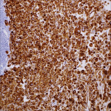
Programmed death-1 (PD-1) is expressed on activated T-cells, B-cells, and myeloid cells. Anti-PD-1 is a marker of angioimmunoblastic lymphoma and suggests a unique cell of origin for this neoplasm. Unlike CD10 and BCL6, PD-1 is expressed by few B-cells, so anti-PD-1 may be a more specific and useful diagnostic marker in angioimmunoblastic lymphoma. In addition, PD-1 expression provides evidence that angioimmunoblastic lymphoma is a neoplasm derived from germinal center-associated T-cells. PD-1 expression in angioimmunoblastic lymphoma lends further support to this model of T-cell oncogenesis, in which specific subtypes of T-cells may undergo neoplastic transformation and result in specific distinct histologic, immunophenotypic, and clinical subtypes of T-cell neoplasia.
Programmed death-1 (PD-1), also known as CD279, is a type-I transmembrane protein expressed on T-cells, B-cells, and monocytes during activation. PD-1 and its ligands (PD-L1 and PD-L2) function as an immune checkpoint pathway by regulating T-cell activation and autoimmunity. PD-1 labels follicular helper T-cells and is therefore a useful marker for angioimmunoblastic T-cell lymphoma.

Quality


IVD

IVD

IVD

RUO

Linkage

PD-1 Positive Control Slides, Product No. 315S, are available for immunohistochemistry (formalin-fixed, paraffin-embedded sections).

Physical form

Solution in Tris Buffer, pH 7.3-7.7, with 1% BSA and <0.1% Sodium Azide

Preparation Note

Download the IFU specific to your product lot and formatNote: This requires a keycode which can be found on your packaging or product label.

Other Notes

For Technical Service please contact: 800-665-7284 or email: service@cellmarque.com

Legal Information

Cell Marque is a trademark of Merck KGaA, Darmstadt, Germany

Not finding the right product?  

Try our Product Selector Tool.


Certificates of Analysis (COA)

Search for Certificates of Analysis (COA) by entering the products Lot/Batch Number. Lot and Batch Numbers can be found on a product’s label following the words ‘Lot’ or ‘Batch’.

Already Own This Product?

Find documentation for the products that you have recently purchased in the Document Library.

Visit the Document Library

Norikazu Mataki et al.
The American journal of gastroenterology, 102(2), 302-312 (2007-02-22)
PD-L1 (also B7-H1) and PD-L2 (also B7-DC) are ligands for programmed death-1 (PD-1), which is a member of the CD28/B7 superfamily of costimulatory molecules and plays an inhibitory role on the periphery. Impaired regulation of this system may cause disruption
David M Dorfman et al.
The American journal of surgical pathology, 30(7), 802-810 (2006-07-05)
Programmed death-1 (PD-1), a member of the CD28 costimulatory receptor family, is expressed by germinal center-associated T cells in reactive lymphoid tissue. In a study of a wide range of lymphoproliferative disorders, neoplastic T cells in 23 cases of angioimmunoblastic
Junzo Hamanishi et al.
Proceedings of the National Academy of Sciences of the United States of America, 104(9), 3360-3365 (2007-03-16)
The ligands for programmed cell death 1 (PD-1), an immunoinhibitory receptor belonging to CD28/cytotoxic T lymphocyte antigen 4 family, are PD-1 ligand 1 and 2 (PD-Ls). Recent reports suggest that the aberrant expression of PD-Ls on tumor cells impairs antitumor
Anne Isine Bolstad et al.
Arthritis and rheumatism, 48(1), 174-185 (2003-01-16)
To assess salivary gland tissues obtained from patients with primary Sjögren's syndrome (SS) for the gene expression profile of the candidate genes TNFRSF6 (Fas), TNFSF6 (FasL), SSA1 (Ro52 alpha and the splice variant Ro52 beta), SSB (La), CTLA4, PDCD1 (PD-1)
Masaya Kobayashi et al.
The Journal of rheumatology, 32(11), 2156-2163 (2005-11-03)
Programmed death-1 (PD-1) mediates a negative signal and introduces tolerance for lymphocytes. Dysfunction of the PD-1 pathway is thought to result in autoimmune diseases such as rheumatoid arthritis (RA). To investigate the role of the PD-1/PD-L system in the pathology

Our team of scientists has experience in all areas of research including Life Science, Material Science, Chemical Synthesis, Chromatography, Analytical and many others.

Contact Technical Service