Skip to Content
MilliporeSigma
All Photos(1)

Documents

383A-7

Sigma-Aldrich

SOX-10 Rabbit Polyclonal Antibody

Sign Into View Organizational & Contract Pricing


About This Item

UNSPSC Code:
12352200
NACRES:
NA.41

biological source

rabbit

Quality Level

100
500

conjugate

unconjugated

antibody form

Ig fraction of antiserum

antibody product type

primary antibodies

clone

polyclonal

description

For In Vitro Diagnostic Use in Select Regions (See Chart)

form

buffered aqueous solution

species reactivity

human

packaging

vial of 0.1 mL concentrate (383A-74)
vial of 0.5 mL concentrate (383A-75)
bottle of 1.0 mL predilute (383A-77)
vial of 1.0 mL concentrate (383A-76)
bottle of 7.0 mL predilute (383A-78)

manufacturer/tradename

Cell Marque

technique(s)

immunohistochemistry (formalin-fixed, paraffin-embedded sections): 1:25-1:100

control

melanoma, schwannoma, skin melanocytes

shipped in

wet ice

storage temp.

2-8°C

visualization

nuclear

Gene Information

human ... SOX10(6663)
mouse ... Sox10(20665)

General description

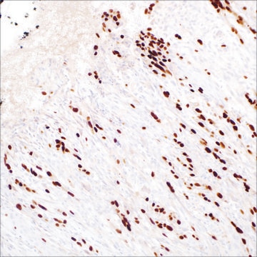
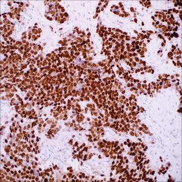
Sry-related HMG-BOX gene 10, (SOX-10), a nuclear transcription factor that participates in neural crest development and in the specification and differentiation of cells of melanocytic lineage, has been recently shown to be a sensitive marker of melanoma, including conventional, spindled, and desmoplastic subtypes. SOX-10 nuclear expression was found in 76 of 78 melanomas (97%) and 38 of 77 malignant peripheral nerve sheath tumors (49%), whereas S100 protein was expressed in 71 melanomas (91%) and 23 malignant peripheral nerve sheath tumors (30%). SOX-10 was expressed by metastatic melanomas and nodal capsular nevus in sentinel lymph nodes, but not by other lymph node components such as dendritic cells which usually express S100 protein. It is known that the commonly used melanoma markers, anti-HMB-45 and anti-Melan-A, are poorly expressed in desmoplastic melanomas while it has been reported that anti-SOX-10 was moderately to strongly expressed in all of the 28 desmoplastic melanomas tested. SOX-10 is less likely expressed by background fibrocytes and histiocytes in exicion scar than S100 and MiTF. Therefore, anti-SOX-10 is considered a very reliable marker for recognizing residual demoplastic melanoma.

SOX-10 is diffusely expressed in schwannomas and neurofibromas. SOX-10 reaction is not identified in any other mesenchymal and epithelial tumors except for myoepitheliomas and diffuse astrocytomas. SOX-10 expression is seen in sustentacular cells of pheochromocytomas and paragangliomas, and occasionally carcinoid tumors from various organs, but not in the tumor cells.

In normal tissues, SOX-10 is expressed in Schwann cells, melanocytes, and myoepithelial cells of salivary, bronchial, eccrine, and mammary glands. SOX-10 expression is also observed in mast cells in a variety of tissues and organs in both nuclear and cytoplasmic reaction.

Quality


IVD

IVD

IVD

RUO

Linkage

SOX-10 Positive Control Slides, Product No. 383S, are available for immunohistochemistry (formalin-fixed, paraffin-embedded sections).

Physical form

Solution in Tris Buffer, pH 7.3-7.7, with 1% BSA and <0.1% Sodium Azide

Preparation Note

Download the IFU specific to your product lot and formatNote: This requires a keycode which can be found on your packaging or product label.

Other Notes

For Technical Service please contact: 800-665-7284 or email: service@cellmarque.com

Legal Information

Cell Marque is a trademark of Merck KGaA, Darmstadt, Germany

Still not finding the right product?  

Give our Product Selector Tool a try.

Storage Class

12 - Non Combustible Liquids

wgk_germany

WGK 2

flash_point_f

Not applicable

flash_point_c

Not applicable


Certificates of Analysis (COA)

Search for Certificates of Analysis (COA) by entering the products Lot/Batch Number. Lot and Batch Numbers can be found on a product’s label following the words ‘Lot’ or ‘Batch’.

Already Own This Product?

Find documentation for the products that you have recently purchased in the Document Library.

Visit the Document Library

A Robson et al.
Histopathology, 38(2), 135-140 (2001-02-24)
The histological distinction of desmoplastic melanoma from cutaneous scar tissue, particularly in the context of re-excision specimens or possible recurrence, may be very difficult. Immunostaining for S100 protein is often used to discriminate although there are little data on S100
Joe A Chorny et al.
The American Journal of dermatopathology, 24(4), 309-312 (2002-07-27)
Distinguishing desmoplastic melanoma (DM) from scar tissue on routine microscopy can be difficult, especially in re-excision specimens, and S100 immunohistochemistry has been recommended as a useful adjunct. The purpose of this study is to evaluate the extent and nature of
Robert N Kelsh
BioEssays : news and reviews in molecular, cellular and developmental biology, 28(8), 788-798 (2006-08-24)
For both vertebrate developmental and evolutionary biologists, and also for clinicians, the neural crest (NC) is a fundamental cell population. An understanding of Sox10 function in NC development is of particular significance since Sox10 mutations underlie several neurocristopathies. Surprisingly, experiments
T A Longacre et al.
The American journal of surgical pathology, 20(12), 1489-1500 (1996-12-01)
The clinical, histologic, and immunohistologic features of 22 desmoplastic melanomas (DMM), 10 mixed desmoplastic and spindle-cell melanomas (DMM/SMM), and two cellular spindle-cell melanomas (SMM) were studied. Patients ranged in age from 35 to 91 years (mean, 67) and included 23
Daisuke Nonaka et al.
The American journal of surgical pathology, 32(9), 1291-1298 (2008-07-19)
S100 protein is a sensitive marker for melanomas and peripheral nerve sheath tumors. It is, however, expressed by other mesenchymal and epithelial tumors. Despite its low specificity, S100 protein is valuable for the diagnosis of desmoplastic melanomas and peripheral nerve

Articles

Immunohistochemistry (IHC) techniques and applications have greatly improved, dermatopathology is still largely based on H&E stained slides.This paper outlines ways in which IHC antibodies can be utilized for dermatopathology.

Our team of scientists has experience in all areas of research including Life Science, Material Science, Chemical Synthesis, Chromatography, Analytical and many others.

Contact Technical Service