Skip to Content
MilliporeSigma
All Photos(4)

Documents

G2781

Sigma-Aldrich

Anti-Glutamine Synthetase antibody produced in rabbit

IgG fraction of antiserum, buffered aqueous solution

Synonym(s):

Glutamine Synthetase Antibody, Glutamine Synthetase Antibody - Anti-Glutamine Synthetase antibody produced in rabbit

Sign Into View Organizational & Contract Pricing


About This Item

MDL number:
UNSPSC Code:
12352203
NACRES:
NA.41

biological source

rabbit

Quality Level

conjugate

unconjugated

antibody form

IgG fraction of antiserum

antibody product type

primary antibodies

clone

polyclonal

form

buffered aqueous solution

mol wt

antigen 45 kDa

species reactivity

rat

packaging

antibody small pack of 25 μL

technique(s)

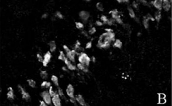
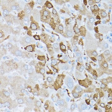
immunohistochemistry (formalin-fixed, paraffin-embedded sections): 1:10,000 using rat brain sections
microarray: suitable
western blot: 1:10,000 using rat brain cytosolic fraction

UniProt accession no.

storage temp.

−20°C

target post-translational modification

unmodified

Gene Information

human ... GLUL(2752)
mouse ... Glul(14645)
rat ... Glul(24957)

General description

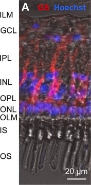
Anti-Glutamine Synthetase is produced in rabbit using a synthetic peptide corresponding to the C-terminus of mouse glutamine synthetase (GS) conjugated to keyhole limpet hemocyanin (KLH) as immunogen. This sequence is identical in human, bovine, rat, hamster and pig GS, and highly conserved in chicken GS. GS is found in mammals as an octamer of identical 45 kDa subunits. It is localized in glial cells of various species. It is present in the brain primarily in astrocytes and in the retina in Muller glial cells that support neurons using glutamate as a neurotransmitter. GS is found in several brain regions including hippocampus, cerebral cortex, neostriatum and cerebellar granular layer, corresponding to sites with high glutamate receptor density.

Specificity

Anti-Glutamine synthetase recognizes rat glutamine synthetase (GS) (45 kDa). Staining of GS in immunoblotting is specifically inhibited with the GS immunizing peptide (amino acids 357-373 with Nterminally added lysine).

Immunogen

synthetic peptide corresponding to the C-terminal region of mouse glutamine synthetase (GS) (amino acids 357-373. This sequence is identical in human, bovine, rat, hamster, and pig GS and highly conserved chicken
Gs (single amino acid substitution).

Application


Immunoblotting: a minimum working antibody dilution of 1:10,000 is determined using a rat brain cytosolic fraction extract.
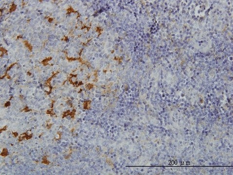
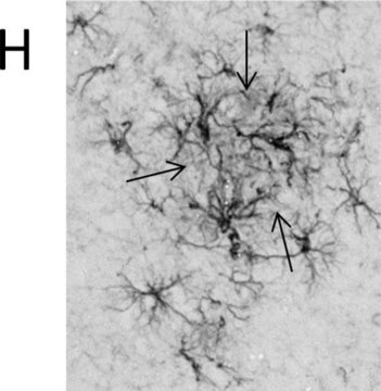
Immunohistochemistry: a minimum working antibody dilution of 1:10,000 is determined using formalin-fixed, paraffin-embedded sections of rat brain.
Anti-Glutamine Synthetase antibody has been used for the detection and localization of GS by immunoblotting and immunohistochemistry.

Biochem/physiol Actions

Glutamine synthetase (GS) catalyzes the amidation of glutamate to form glutamine. In the brain, GS is considered as a key enzyme, participating in recycling of the excitatory neurotransmitter glutamate released from neurons and termination of the neurotransmitter signal, as well as in ammonia detoxification. GS activity is a useful marker for astrocytes and an important differentiation feature in the retina. Upregulation of GS is a hallmark of reactive astrocytosis and GS activity level increases following spinal cord injury. The GS expression in astrocytes is diminished in brains affected by Alzheimer′s disease, particularly in the vicinity of senile plaques. GS is upregulated in a subset of human hepatocellular carcinomas, suggesting that its expression may be related to tumor recurrence, since tumors expressing GS are independent of the extracellular glutamine supply.

Physical form

Solution in 0.01 M phosphate buffered saline, pH 7.4, containing 15 mM sodium azide

Disclaimer

Unless otherwise stated in our catalog or other company documentation accompanying the product(s), our products are intended for research use only and are not to be used for any other purpose, which includes but is not limited to, unauthorized commercial uses, in vitro diagnostic uses, ex vivo or in vivo therapeutic uses or any type of consumption or application to humans or animals.

Not finding the right product?  

Try our Product Selector Tool.

wgk_germany

WGK 2

flash_point_f

Not applicable

flash_point_c

Not applicable


Certificates of Analysis (COA)

Search for Certificates of Analysis (COA) by entering the products Lot/Batch Number. Lot and Batch Numbers can be found on a product’s label following the words ‘Lot’ or ‘Batch’.

Already Own This Product?

Find documentation for the products that you have recently purchased in the Document Library.

Visit the Document Library

Effects of bevacizumab (Avastin) on retinal cells in organotypic culture
Kaempf S, et al.
Investigative Ophthalmology & Visual Science, 49(7), 3164-3171 (2008)
Deniz Dalkara et al.
Molecular therapy : the journal of the American Society of Gene Therapy, 19(9), 1602-1608 (2011-04-28)
Mutations in over 80 identified genes can induce apoptosis in photoreceptors, resulting in blindness with a prevalence of 1 in 3,000 individuals. This broad genetic heterogeneity of disease impacting a wide range of photoreceptor functions renders the design of gene-specific
Ying Zhu et al.
mBio, 8(4) (2017-08-16)
While glutamine is a nonessential amino acid that can be synthesized from glucose, some cancer cells primarily depend on glutamine for their growth, proliferation, and survival. Numerous types of cancer also depend on asparagine for cell proliferation. The underlying mechanisms
Peter X Shaw et al.
Investigative ophthalmology & visual science, 57(11), 5083-5092 (2016-09-30)
We have previously demonstrated that soluble adenylyl cyclase (sAC) is necessary for retinal ganglion cell (RGC) survival and axon growth. Here, we further investigate the role of sAC in neuronal differentiation during retinal development. Chx10 or Math5 promoter-driven Cre-Lox recombination
Ala Moshiri et al.
Investigative ophthalmology & visual science, 58(3), 1709-1718 (2017-03-23)
Small guanosine triphosphatase (GTPase) ADP-ribosylation factors (Arfs) regulate membrane traffic and actin reorganization under the control of GTPase-activating proteins (GAPs). Arap1 is an Arf-directed GAP that inhibits the trafficking of epidermal growth factor receptor (EGFR) to the early endosome, but

Our team of scientists has experience in all areas of research including Life Science, Material Science, Chemical Synthesis, Chromatography, Analytical and many others.

Contact Technical Service