SAE0151
Proteinase K from Tritirachium album
free of DNA contaminants, suitable for Microbiome research, lyophilized powder, ≥30 units/mg protein
Synonym(s):
Proteinase K from Tritirachium album, Endopeptidase K
About This Item
Recommended Products
Quality Level
specific activity
30 units/mg protein
mol wt
28.93 kDa
feature
DNA free
shipped in
wet ice
storage temp.
−20°C
Looking for similar products? Visit Product Comparison Guide
Related Categories
General description
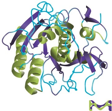
Proteinase K is a stable serine protease with broad substrate specificity. It degrades many proteins in the native state even in the presence of detergents. Proteinase K was isolated from a fungus able to grow on keratin and the enzyme can digest native keratin. The enzyme belongs to the subtilisin family with an active site catalytic triad (Asp39 -His69 -Ser224).
The predominant site of cleavage is the peptide bond adjacent to the carboxyl group of aliphatic and aromatic amino acids with blocked alpha amino groups. It is commonly used for its broad specificity. Proteinase K is usually denatured by subsequent phenol extractions.
The study of microbial communities has been revolutionized in recent years by the widespread adoption of culture independent analytical techniques such as 16S rRNA gene sequencing and metagenomics. Since DNA contamination during sample preparation is a major problem of these sequence-based approaches, DNA extraction reagents free of DNA contaminates are essential.
Application
- Proteinase K is frequently used in molecular biology applications to digest unwanted proteins, such as nucleases from DNA or RNA preparations from microorganisms, cultured cells, and plants.
- Removes endotoxins that bind to cationic proteins such as lysozyme and ribonuclease A useful for the isolation of hepatic, yeast, and mung bean mitochondria.
- Determination of enzyme localization on membranes
- Treatment of paraffin embedded tissue sections to expose antigen binding sites for antibody labeling.
- Digestion of proteins from brain tissue samples for prions in Transmissible Spongiform Encephalopathies (TSE) research.
Removes endotoxins that bind to cationic proteins such as lysozyme and ribonuclease A.
Reported useful for the isolation of hepatic, yeast, and mung bean mitochondria
Determination of enzyme localization on membranes
Treatment of paraffin embedded tissue sections to expose antigen binding sites for antibody labeling.
Digestion of proteins from brain tissue samples for prions in Transmissible Spongiform Encephalopathies (TSE) research.
Biochem/physiol Actions
Features and Benefits
Unit Definition
Preparation Note
signalword
Danger
hcodes
Hazard Classifications
Eye Irrit. 2 - Resp. Sens. 1 - Skin Irrit. 2 - STOT SE 3
target_organs
Respiratory system
wgk_germany
WGK 1
flash_point_f
Not applicable
flash_point_c
Not applicable
Certificates of Analysis (COA)
Search for Certificates of Analysis (COA) by entering the products Lot/Batch Number. Lot and Batch Numbers can be found on a product’s label following the words ‘Lot’ or ‘Batch’.
Already Own This Product?
Find documentation for the products that you have recently purchased in the Document Library.
Articles
Enzymes provide a non-mechanical method for cell lysis and protoplast preparation. It may seem like a simple process to throw in your enzyme, stick your tube in the water-bath and walk away, but what is actually going on in that process?
Our team of scientists has experience in all areas of research including Life Science, Material Science, Chemical Synthesis, Chromatography, Analytical and many others.
Contact Technical Service


