AB5726
Anti-Dlx2 Antibody
Chemicon®, from rabbit
Synonym(s):
Distalless
About This Item
Recommended Products
biological source
rabbit
Quality Level
antibody form
affinity purified immunoglobulin
antibody product type
primary antibodies
clone
polyclonal
purified by
affinity chromatography
species reactivity
mouse, human
manufacturer/tradename
Chemicon®
technique(s)
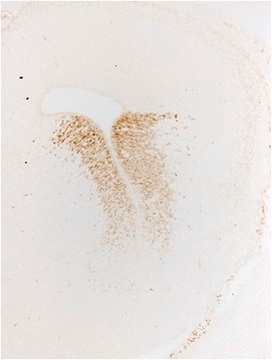
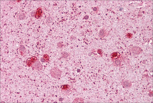
immunohistochemistry: suitable
western blot: suitable
NCBI accession no.
UniProt accession no.
shipped in
dry ice
target post-translational modification
unmodified
Gene Information
human ... DLX2(1746)
Specificity
Immunogen
Application
Neuroscience
Developmental Neuroscience
Neuronal & Glial Markers
Immunohistochemistry: 1:200-1:1,000.
Optimal working dilutions must be determined by the end user.
Physical form
Storage and Stability
PREPARATION AND USE:
To reconstitute the antibody, centrifuge the antibody vial at moderate speed (5,000 rpm) for 5 minutes to pellet the precipitated antibody product. Carefully remove the ammonium sulfate/PBS buffer solution and discard. It is not necessary to remove all of the ammonium sulfate/PBS solution: 10 μL of residual ammonium sulfate solution will not affect the resuspension of the antibody. Do not let the protein pellet dry, as severe loss of antibody reactivity can occur.
Resuspend the antibody pellet in a suitable biological buffer such as PBS or TBS (pH 7.3-7.5) to a final concentration of 1.0 mg/mL. For example, to achieve a 1.0 mg/mL concentration with 50 μg of precipitated antibody, the amount of buffer needed would be 50 μL.
Carefully add the liquid buffer to the pellet. DO NOT VORTEX. Mix by gentle stirring with a wide pipet tip or gentle finger-tapping. Let the precipitated antibody rehydrate for 1 hour at 4-25°C prior to use. Small particles of precipitated antibody that fail to resuspend are normal. Vials are overfilled to compensate for any losses.
The rehydrated antibody solutions can be stored undiluted at 2-8°C for 2 months without any significant loss of activity. Note, the solution is not sterile, thus care should be taken if product is stored at 2-8°C.
For storage at -20°C, the addition of an equal volume of glycerol can be used, however, it is recommended that ACS grade or higher glycerol be used, as significant loss of activity can occur if the glycerol used is not of high quality.
For long-term storage at -70°C, it is recommended that the rehydrated antibody solution be further diluted 1:1 with a 2% BSA (fraction V, highest-grade available) solution made with the rehydration buffer. The resulting 1% BSA/antibody solution can be aliquoted and stored frozen at -70°C for up to 6 months. Avoid repeated freeze/thaw cycles.
Legal Information
Disclaimer
Not finding the right product?
Try our Product Selector Tool.
wgk_germany
WGK 2
flash_point_f
Not applicable
flash_point_c
Not applicable
Certificates of Analysis (COA)
Search for Certificates of Analysis (COA) by entering the products Lot/Batch Number. Lot and Batch Numbers can be found on a product’s label following the words ‘Lot’ or ‘Batch’.
Already Own This Product?
Find documentation for the products that you have recently purchased in the Document Library.
Our team of scientists has experience in all areas of research including Life Science, Material Science, Chemical Synthesis, Chromatography, Analytical and many others.
Contact Technical Service